. 2000;9(3):145-56.
doi: 10.3727/000000001783992650.

An antiprion effect of the anticytoskeletal drug latrunculin A in yeast

Affiliations

An antiprion effect of the anticytoskeletal drug latrunculin A in yeast

P A Bailleul-Winslett et al. Gene Expr. 2000.

Abstract

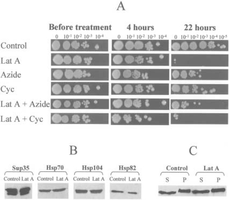
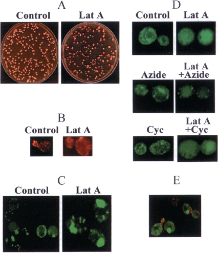
Prions are infectious aggregation-prone isoforms of the normal proteins, supposedly able to seed aggregation of the normal cellular counterparts. In vitro, prion proteins form amyloid fibers, resembling cytoskeletal structures. Yeast prion [PSI], which is a cytoplasmically inherited aggregated isoform of the translation termination factor Sup35p (eRF3), serves as a useful model for studying mechanisms of prion diseases and other amyloidoses. The previously described interaction between Sup35p and cytoskeletal assembly protein Sla1p points to the possible relationships between prions and cytoskeletal networks. Although the Sup35PSI+ aggregates do not colocalize with actin patches, we have shown that yeast cells are efficiently cured of the [PSI] prion by prolonged incubation with latrunculin A, a drug disrupting the actin cytoskeleton. On the other hand, treatments with sodium azide or cycloheximide, agents blocking yeast protein synthesis and cell proliferation but not disrupting the cytoskeleton, do not cause a significant loss of [PSI]. Moreover, simultaneous treatment with sodium azide or cycloheximide blocks [PSI] curing by latrunculin A, indicating that prion loss in the presence of latrunculin A requires a continuation of protein synthesis during cytoskeleton disruption. The sodium azide treatment also decreases the toxic effect of latrunculin A. Latrunculin A influences neither the levels of total cellular Sup35p nor the levels of chaperone proteins, such as Hsp104 and Hsp70, which were previously shown to affect [PSI]. This makes an indirect effect of latrunculin A on [PSI] via induction of Hsps unlikely. Fluorescence microscopy detects changes in the structure and/or localization of the Sup35PSI+ aggregates in latrunculin A-treated cells. We conclude that the stable maintenance of the [PSI] prion aggregates in the protein-synthesizing yeast cells partly depends on an intact actin cytoskeleton, suggesting that anticytoskeletal treatments could be used to counteract some aggregation-related disorders.


Figures

Figure 1
Figure 1
Effects of latrunculin A on yeast viability and protein levels. (A) Viability of yeast cells in the control culture and cultures treated with latrunculin A (Lat A), sodium azide (Azide), and cycloheximide (Cyc) in various combinations. Cultures were grown in YPD at 30°C as described in Materials and Methods. At each time point, serial decimal dilutions were made, and 3-μ1 aliquots of each dilution were spotted onto YPD plates. Plates have been photographed after 3 days of incubation at 30°C. Concentrations: latrunculin A, 200 μM; sodium azide, 10 mM; cycloheximide, 100 μg/ml. (B) Levels of the Sup35p and Hsps in the control (Control) and latrunculin A-treated (Lat A) cultures of the strain OT55, as determined by Western blotting. Latrunculin A treatment was for 4 h. Equal amounts of total cellular protein were loaded in each lane, as verified by Coomassie staining (not shown). (C) Distribution of Sup35p between soluble (S, supernatant) and insoluble (P, pellet) fractions is not affected by latrunculin A. Yeast extracts were prepared and fractionated as described (32). Latrunculin A treatment was for 4 h.
Figure 2
Figure 2
[PSI] loss and patterns of the Sup35p aggregates in the control and latrunculin A-treated cells. (A) Latrunculin A-induced loss of [PSI] as visualized by color on YPD plates. The cultures of the [PSI +] strain OT55 were plated onto YPD medium after 18 h of treatment with latrunculin A (Lat A, right) or without such a treatment (Control, left). Red colonies are [psi ], while light pink colonies are [PSI +]. (B) Latrunculin A causes dissociation of the actin patches. Actin patches were visualized by rhodamine-phalloidin staining (red). Patches are clearly seen in the control cells of the strain OT55 and preferentially concentrate in the area of new bud and bud neck (left). Most patches disappear after latrunculin A treatment; red background indicates that actin becomes distributed evenly in the cytoplasm (right). Latrunculin A treatment was for 4 h. (C) Visualization of the Sup35pPSI+ aggregates in the yeast strain transformed with the plasmid pHGPD-NMsGFP, containing the chimeric SUP35NM-GFP gene under the control of strong GPD promoter. Most of the Sup35NM-GFP aggregates (green) are relatively small and compact in the control cells of the [PSI +] strain OT55 (left). Four-hour treatment with 200 μM latrunculin A leads to diffusion of the aggregates (right). Images of the control and latrunculin A-treated cells were taken at the same scale. Difference in size is due to uncontrolled cell growth in the conditions when cell division is blocked for 4 h by latrunculin A treatment. (D) Visualization of the Sup35pPSI+ aggregates in the yeast strain transformed with the plasmid pLSpSUP35NM-GFP, containing the chimeric SUP35NM-GFP gene under the control of the SUP35 own promoter, which is moderately expressed. Due to lower levels of the Sup35-GFP production, the prion aggregates in the control cells are smaller in size, compared with (C). Sodium azide blocks the Sup35-GFP diffusion caused by latrunculin A more efficiently than cycloheximide. See also comments in the text. All treatments were for 1 h. Similar results were observed after 4-h treatments (not shown). All images were taken at the same scale. Concentrations: latrunculin A, 200 μM; sodium azide, 10 mM; cycloheximide, 100 μg/ml. (E) Actin patches (red) usually do not colocalize with the Sup35NM-GFP aggregates (green) in the cells of [PSI +] strain OT55. The actin patches were visualized by rhodamine-phalloidin staining as in (B). The fluorescent Sup35-GFP protein was produced by the plasmid pHGPD-NMsGFP, as in (C). Images used for (B)–(E) were obtained by using a confocal microscope, as described in Materials and Methods.

Similar articles

Cited by

References

    1. Adams A. E. M.; Pringle J. Relationship of actin and tubulin distribution to bud growth in wild-type and morphogenetic mutant of Saccharomyces cerevisiae . J. Cell. Biol. 98:934–945; 1984. - PMC - PubMed
    1. Ayscough K. Use of latrunculin-A, an actin monomer-binding drug. Methods Enzymol. 298:18–25; 1998. - PubMed
    1. Ayscough K. R.; Stryker J.; Pokala N.; Sanders M.; Crews P.; Drubin D. G. High rates of actin filament turnover in budding yeast and roles for actin in establishment and maintenance of cell polarity revealed using the actin inhibitor latrunculin-A. J. Cell Biol. 137: 399–416; 1997. - PMC - PubMed
    1. Bailleul P. A.; Newnam G. P.; Steenbergen J. N.; Chernoff Y. O. Genetic study of interactions between the cytoskeletal assembly protein Slal and prion-forming domain of the release factor Sup35 (eRF3) in Saccharomyces cerevisiae . Genetics 153:81–94; 1999. - PMC - PubMed
    1. Broach J. R.; Strathern J. N.; Hicks J. B. Transformation of yeast: Development of the hybrid cloning vector and isolation of the CAN1 gene. Gene 8:121–133; 1979. - PubMed

Publication types

MeSH terms

LinkOut - more resources