. 2001 Apr;183(7):2204-11.
doi: 10.1128/JB.183.7.2204-2211.2001.

Genetic evidence that the alpha5 helix of the receiver domain of PhoB is involved in interdomain interactions

Affiliations

Genetic evidence that the alpha5 helix of the receiver domain of PhoB is involved in interdomain interactions

M P Allen et al. J Bacteriol. 2001 Apr.

Abstract

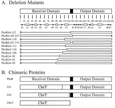
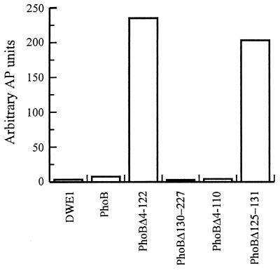
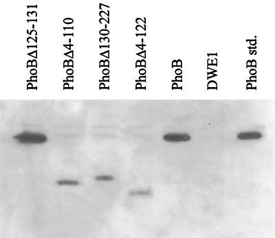
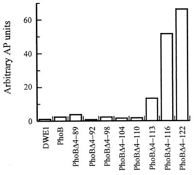
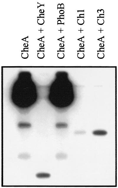
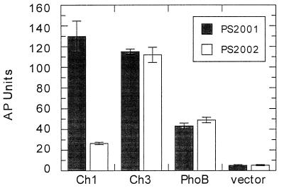
Two-component signaling proteins are involved in transducing environmental stimuli into intracellular signals. Information is transmitted through a phosphorylation cascade that consists of a histidine protein kinase and a response regulator protein. Generally, response regulators are made up of a receiver domain and an output domain. Phosphorylation of the receiver domain modulates the activity of the output domain. The mechanisms by which receiver domains control the activities of their respective output domains are unknown. To address this question for the PhoB protein from Escherichia coli, we have employed two separate genetic approaches, deletion analysis and domain swapping. In-frame deletions were generated within the phoB gene, and the phenotypes of the mutants were analyzed. The output domain, by itself, retained significant ability to activate transcription of the phoA gene. However, another deletion mutant that contained the C-terminal alpha-helix of the receiver domain (alpha5) in addition to the entire output domain was unable to activate transcription of phoA. This result suggests that the alpha5 helix of the receiver domain interacts with and inhibits the output domain. We also constructed two chimeric proteins that join various parts of the chemotaxis response regulator, CheY, to PhoB. A chimera that joins the N-terminal approximately 85% of CheY's receiver domain to the beta5-alpha5 loop of PhoB's receiver domain displayed phosphorylation-dependent activity. The results from both sets of experiments suggest that the regulation of PhoB involves the phosphorylation-mediated modulation of inhibitory contacts between the alpha5 helix of its unphosphorylated receiver domain and its output domain.


Figures

FIG. 1
FIG. 1
Structures of the deletion and chimeric proteins used in this study. (A) The domain structure of PhoB is represented as two white rectangles separated by a black linker region. The amino acid numbers are shown above the map of the secondary structures of PhoB (arrows, β-strands; ovals, α-helices) (30, 35). For the 10 deletion proteins the white bars represent the protein segments that remain whereas the lines correspond to the deleted segments. The name of each protein designates which amino acid residues have been deleted from PhoB. For example, PhoBΔ4-122 contains residues 1 to 3 of PhoB, followed by Gly-Ser (from an inserted BamHI site in the coding sequence), followed by residues 123 to 229. (B) Schematic representation of the chimeric proteins used in this study. Ch1 joins the N-terminal 108 residues of CheY to the C-terminal 125 residues of PhoB. Ch3 joins the N-terminal 127 amino acid residues of CheY to the C-terminal 106 residues of PhoB.
FIG. 2
FIG. 2
The transcriptional activation activities of PhoB deletion proteins were determined by measuring the amounts of alkaline phosphatase (AP) synthesized in phosphate-sufficient medium. E. coli DWE1 cells were transformed with plasmids encoding PhoB deletion proteins. The genes for PhoB, PhoBΔ4-122, PhoBΔ130-227, PhoBΔ4-110, and PhoBΔ125-131 were contained on plasmids pDE1, pMP40, pMP7, pMP8, and pMP40, respectively. The cells were grown overnight in LB medium containing ampicillin, and alkaline phosphatase assays were performed.
FIG. 3
FIG. 3
Western immunoblot analysis of PhoB, PhoBΔ4-122, PhoBΔ130-227, PhoBΔ4-110, and PhoBΔ125-131. E. coli DWE1 cells were transformed with plasmids encoding PhoB and the four deletion mutants. The cells were grown overnight in LB medium containing ampicillin, were collected by centrifugation, and were lysed in SDS-PAGE sample buffer. Equal amounts of cellular extracts were separated by SDS-PAGE, transferred onto a nitrocellulose membrane, and detected with a chemiluminescence detection system using rabbit anti-PhoB polyclonal sera. Purified PhoB was run as a standard (48).
FIG. 4
FIG. 4
The transcriptional activation activities of a series of PhoB deletion proteins localize the inhibitory region of the receiver domain to the α5 helix. E. coli DWE1 cells were transformed with plasmids encoding PhoB deletion proteins. The genes for PhoB, PhoBΔ4-89, PhoBΔ4-92, PhoBΔ4-98, PhoBΔ4-104, PhoBΔ4-110, PhoBΔ4-113, PhoBΔ4-116, and PhoBΔ4-122 were contained on plasmids pDE1, pMP41, pMP42, pMP44, pMP46, pMP8, pMP48, pMP49, and pMP40, respectively. The cells were grown overnight in glucose-MOPS minimal medium containing 5.0 mM KH2PO4, and alkaline phosphatase (AP) assays were performed.
FIG. 5
FIG. 5
Phosphotransfer reactions between CheA and chimeric proteins Ch1 and Ch3. CheA was phosphorylated with [γ-32P]ATP. Aliquots of [γ-32P]phospho-CheA were mixed with the indicated phosphoacceptors, incubated for 2 min at room temperature, and analyzed by SDS-PAGE and autoradiography. The final concentrations of the phosphoacceptors in the phosphotransfer reactions were as follows: CheY, 12 μM; PhoB, 15 μM; Ch1, 2 μM; Ch3, 12 μM.
FIG. 6
FIG. 6
Alkaline phosphatase (AP) assay to measure the output activities of chimeric proteins Ch1 and Ch3. The genes for Ch1, Ch3, and PhoB were cloned into vector pMLB1120.215 and transformed into either PS2001 or PS2002. The data show means and standard deviations of assays performed in triplicate.

References

    1. Alon U, Camarena L, Surette M G, Arcas B A, Liu Y, Leibler S, Stock J B. Response regulator output in bacterial chemotaxis. EMBO J. 1998;17:4238–4248. - PMC - PubMed
    1. Baikalov I, Schroder I, Kaczor-Grzeskowiak M, Grzeskowiak K, Gunsalus R P, Dickerson R E. Structure of the Escherichia coli response regulator NarL. Biochemistry. 1996;35:11053–11061. - PubMed
    1. Birck C, Mourey L, Gouet P, Fabry B, Schumacker J, Rousseau P, Kahn D, Samama J-P. Conformational changes induced by phosphorylation of the FixJ receiver domain. Structure. 1999;7:1505–1515. - PubMed
    1. Cho H S, Lee S Y, Yan D, Pan X, Parkinson J S, Kustu S, Wemmer D E, Pelton J G. NMR structure of activated CheY. J Mol Biol. 2000;297:543–551. - PubMed
    1. Djordjevic S, Goudreau P N, Xu Q, Stock A M, West A H. Structural basis for methylesterase CheB regulation by a phosphorylation-activated domain. Proc Natl Acad Sci USA. 1998;95:1381–1386. - PMC - PubMed

Publication types

LinkOut - more resources