. 2001 Mar 15;21(6):1975-82.
doi: 10.1523/JNEUROSCI.21-06-01975.2001.

Extracellular ATP or ADP induce chemotaxis of cultured microglia through Gi/o-coupled P2Y receptors

Affiliations

Extracellular ATP or ADP induce chemotaxis of cultured microglia through Gi/o-coupled P2Y receptors

S Honda et al. J Neurosci. .

Abstract

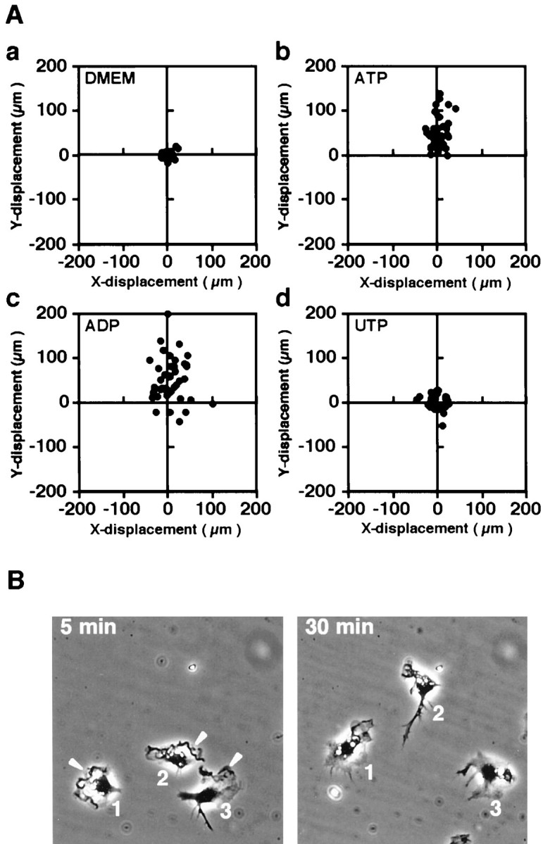
The initial microglial responses that occur after brain injury and in various neurological diseases are characterized by microglial accumulation in the affected sites of brain that results from the migration and proliferation of these cells. The early-phase signal responsible for this accumulation is likely to be transduced by rapidly diffusible factors. In this study, the possibility of ATP released from injured neurons and nerve terminals affecting cell motility was determined in rat primary cultured microglia. Extracellular ATP and ADP induced membrane ruffling and markedly enhanced chemokinesis in Boyden chamber assay. Further analyses using the Dunn chemotaxis chamber assay, which allows direct observation of cell movement, revealed that both ATP and ADP induced chemotaxis of microglia. The elimination of extracellular calcium or treatment with pyridoxalphosphate-6-azophenyl-2',4'-disulphonic acid, suramin, or adenosine-3'-phosphate-5'-phosphosulfate did not inhibit ATP- or ADP-induced membrane ruffling, whereas AR-C69931MX or pertussis toxin treatments clearly did so. As an intracellular signaling molecule underlying these phenomena, the small G-protein Rac was activated by ATP and ADP stimulation, and its activation was also inhibited by pretreatment with pertussis toxin. These results strongly suggest that membrane ruffling and chemotaxis of microglia induced by ATP or ADP are mediated by G(i/o)-coupled P2Y receptors.


Figures

Fig. 1.
Fig. 1.
Nucleotide-induced membrane ruffling in microglia. The cells were stimulated with PBS (A) or 50 μm ATP (B), ADP (C), or UTP (D) for 5 min. After fixation, the cells were stained with Texas Red-conjugated phalloidin. ATP and ADP clearly induced membrane ruffling (indicated byarrows). Scale bar, 20 μm.
Fig. 2.
Fig. 2.
Nucleotide-induced chemokinesis of microglia in the Boyden chamber. The cells were exposed to ATP (circle), ADP (square), or UTP (triangle) for 90 min. Nucleotides were added to both top and bottom wells at the concentrations indicated. The absorbance of the stained cells on the bottom side of the filter was measured with a plate reader. Each point and vertical linerepresent the mean and SD for three wells. We confirmed that the three independent experiments showed the same tendency.
Fig. 3.
Fig. 3.
Nucleotide-induced chemotaxis of microglia in the Dunn chemotaxis chamber. A, Vector diagrams of cell displacement at 60 min after setting up the chamber. The cells were incubated in the absence of nucleotides (a) or in the presence of 50 μm ATP (b), ADP (c), or UTP (d) in the outer well. The position of the outer well of the chamber is vertically upward. All diagrams were obtained from a representative experiment using the same lot of microglial culture. We confirmed that the three independent experiments showed the same tendency. B,Displacement and morphological change at 5 and 30 min after setting up the chamber. The cells were incubated in the presence of 50 μm ATP in the outer well. The images of the same area are presented such that the position of the outer well of the chamber is vertically upward. Arrowheads indicate membrane ruffles.
Fig. 4.
Fig. 4.
Effect of extracellular calcium deprivation on membrane ruffling induced by ATP or ADP. Cells were stimulated with PBS (A, D), 50 μm ATP (B, E), or 50 μm ADP (C, F) in BSS with (A–C) or without (D–F) calcium. After stimulation, the cells were fixed and stained with Texas Red-conjugated phalloidin. Arrowheads indicate membrane ruffles. Scale bar, 20 μm.
Fig. 5.
Fig. 5.
Effects of PPADS, suramin, and A3P5PS on nucleotide-induced membrane ruffling. Microglia were stimulated with 50 μm ATP (B, E, H, K) or ADP (C, F, I, L) for 5 min after 10 min of pretreatment with PBS (A–C), 300 μm suramin (D–F), 300 μm PPADS (G–I), or 300 μm A3P5PS (J–L). After fixation, the cells were stained with Texas Red-conjugated phalloidin. Arrowheadsindicate membrane ruffles. Scale bar, 20 μm.
Fig. 6.
Fig. 6.
Effect of AR-C69931MX on nucleotide-induced membrane ruffling. Microglia were stimulated with 50 μmATP (B, F) or ADP (C, G) or 100 ng/ml M-CSF (D, H) for 5 min after 10 min of pretreatment with PBS (A–D) or 1 μm AR-C69931MX (E–H). After fixation, the cells were stained with Texas Red-conjugated phalloidin.Arrowheads indicate membrane ruffling. Scale bar, 20 μm.
Fig. 7.
Fig. 7.
Effects of PTx on nucleotide-induced membrane ruffling. Microglia were stimulated with PBS (A, E), 50 μm ATP (B, F), 50 μmADP (C, G), or 100 ng/ml M-CSF (D, H) for 5 min after 4 hr of pretreatment with PBS (A–D) or 50 ng/ml pertussis toxin (E–H). After fixation, the cells were stained with Texas Red-conjugated phalloidin. Arrowheadsindicate membrane ruffles. Scale bar, 20 μm.
Fig. 8.
Fig. 8.
Effect of PTx on ATP-induced chemokinesis in the Boyden chamber. The chemokinesis assay was performed in the presence of 50 μm ATP with (white column) or without (black column) pertussis toxin treatment. Cells were pretreated with 50 ng/ml PTx for 4 hr.
Fig. 9.
Fig. 9.
Rac activation induced by ATP and ADP.A, Translocation of Rac. The cells were stimulated with PBS (a–c), 50 μm ATP (d–f), or 50 μm ADP (g–i) for 5 min and stained with Rac antibody and FITC-conjugated anti-mouse IgG (a, d, g) and Texas Red-conjugated phalloidin (b, e, h). Merged photographs show the colocalization of Rac and F-actin in the ruffling region (f, i), as indicated by arrows. Scale bar, 10 μm. B, Pull-down assay of activated Rac. Cells were stimulated with PBS (control), 50 μm ATP, or 50 μm ADP for 1 min, and pull-down assay was performed as described in Materials and Methods. Although the total amount of Rac in the cell lysate was the same for each stimulation, activated Rac was increased in the pull-down samples from ATP- and ADP-stimulated cells.
Fig. 10.
Fig. 10.
Effect of PTx on the nucleotide-induced activation of Rac. Pull-down assay was performed, as described in Materials and Methods. Microglia were stimulated with PBS (control), 50 μm ADP, 50 μm ATP, or 100 ng/ml M-CSF for 1 min after 4 hr of treatment with (+) or without (−) PTx at 50 ng/ml.

References

    1. Alexander S, Peters J, Mead A, Lewis S. Receptor & ion channel nomenclature supplement, Ed 10, Trends Pharmacol Sci, pp 64–69. Elsevier Science; London: 1999.
    1. Baltensperger K, Porzig H. The P2U purinoceptor obligatorily engages the heterotrimeric G protein G16 to mobilize intracellular Ca2+ in human erythroleukemia cells. J Biol Chem. 1997;272:10151–10159. - PubMed
    1. Barnard EA, Simon J, Webb TE. Nucleotide receptors in the nervous system. An abundant component using diverse transduction mechanisms. Mol Neurobiol. 1997;15:103–129. - PubMed
    1. Bokoch GM, Bohl BP, Chuang TH. Guanine nucleotide exchange regulates membrane translocation of Rac/Rho GTP-binding proteins. J Biol Chem. 1994;269:31674–31679. - PubMed
    1. Boyer JL, Romero-Avila T, Schachter JB, Harden TK. Identification of competitive antagonists of P2Y1 receptor. Mol Pharmacol. 1996;50:1323–1329. - PubMed

Publication types

MeSH terms

LinkOut - more resources