

Review
doi: 10.1073/pnas.061025898.
Closing down on glyphosate inhibition--with a new structure for drug discovery
Affiliations
- PMID: 11248008
- PMCID: PMC33334
- DOI: 10.1073/pnas.061025898
Item in Clipboard
Review
Closing down on glyphosate inhibition--with a new structure for drug discovery
Proc Natl Acad Sci U S A.
.
No abstract available
Figures
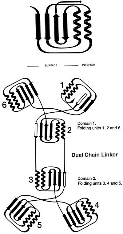
Folding and topological symmetry of EPSPS (adapted from ref. 11).
The two domain structure is formed by 6-fold replication of a protein
folding unit (Upper) comprising two parallel helices and
a four-stranded β-sheet. Each domain is formed from three of these
folding units, which are related by an approximate 3-fold topological
symmetry axis. In the open form of EPSPS, these axes are not collinear,
but are presumably more so in the closed formed of the enzyme reported
by Schönbrunn et al. (10). In each domain, three
of the helices are buried and the surface of the molecule formed from
the three β-sheets and the solvent-accessible faces of the other
three helices. The N and C termini are located in Domain 1 with two
crossover polypeptide segments creating a double hinge that links the
two domains (Lower). Among the six folding units, three
are folded from continuous segments of polypeptide chain. The other
three contain the same arrangement of secondary structural features,
but the sequences are not from a continuous chain. The arrangement
positions the N termini of all 12 helices near the interface that
results when the enzyme closes to form the active site. The positive
macrodipoles from these helices presumably contribute to binding of
substrates, products, and inhibitors, which are all multiply charged
anions.
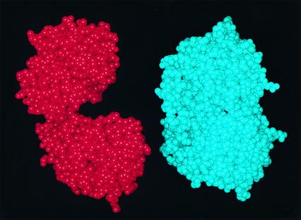
(Left) Space-filling model of E. coli
EPSPS in the open form. (Right) Model of an EPSPS
molecule in the closed, ligand-bound form. Comparisons of the figures
illustrate the dramatic conformational change that attends ligand
binding.
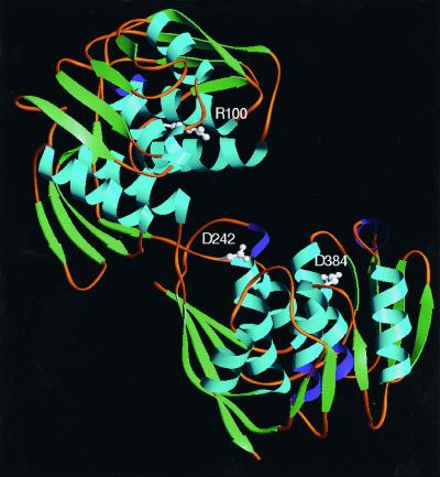
Crystal structure of the open form of EPSPS. Schönbrunn et
al. (10) identify residues in MurA and their homologs in
EPSPS that are determinants in the control of domain closure, and
suggest that inhibitors that bind to these residues will interfere
with closure of the enzymes and the formation of their active sites.
Arg-100 (Upper, or Domain 2) is relatively close to the
active site. Asp-384 is in the Lower domain (Domain 1).
Asp-242 is near the two-stranded hinge that links the two domains.
Comment on
-
Structural basis for the interaction of the fluorescence probe 8-anilino-1-naphthalene sulfonate (ANS) with the antibiotic target MurA.Proc Natl Acad Sci U S A. 2000 Jun 6;97(12):6345-9. doi: 10.1073/pnas.120120397. Proc Natl Acad Sci U S A. 2000. PMID: 10823915 Free PMC article.
-
Interaction of the herbicide glyphosate with its target enzyme 5-enolpyruvylshikimate 3-phosphate synthase in atomic detail.Proc Natl Acad Sci U S A. 2001 Feb 13;98(4):1376-80. doi: 10.1073/pnas.98.4.1376. Proc Natl Acad Sci U S A. 2001. PMID: 11171958 Free PMC article.
-
Structure and topological symmetry of the glyphosate target 5-enolpyruvylshikimate-3-phosphate synthase: a distinctive protein fold.Proc Natl Acad Sci U S A. 1991 Jun 1;88(11):5046-50. doi: 10.1073/pnas.88.11.5046. Proc Natl Acad Sci U S A. 1991. PMID: 11607190 Free PMC article.
References
-
- Baird D D, Upchurch R P, Homesley W B, Franz J E. Proc North Cent Weed Control Conf. 1971;26:64–68.
-
- Franz J E, Mao M K, Sikorski J A. Glyphosate: A Unique Global Herbicide. Washington, DC: Am. Chem. Soc.; 1997.
-
- Jaworski E G. J Agric Food Chem. 1972;20:1195–1198.
-
- Haslam E. Shikimic Acid: Metabolism and Metabolites. New York: Wiley; 1993.
Publication types
MeSH terms
Substances
LinkOut - more resources
Full Text Sources
