. 2001 Mar 13;98(6):2995-3000.
doi: 10.1073/pnas.051520298. Epub 2001 Feb 27.

Structure of a flavin-binding plant photoreceptor domain: insights into light-mediated signal transduction

Affiliations

Structure of a flavin-binding plant photoreceptor domain: insights into light-mediated signal transduction

S Crosson et al. Proc Natl Acad Sci U S A. .

Abstract

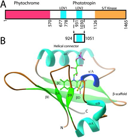
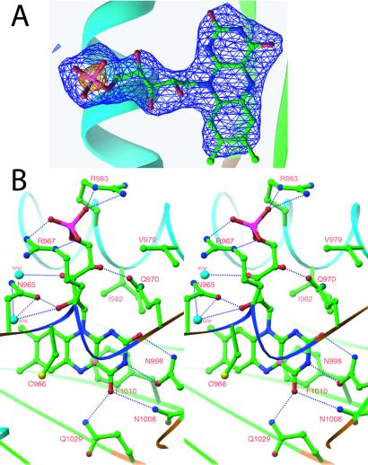
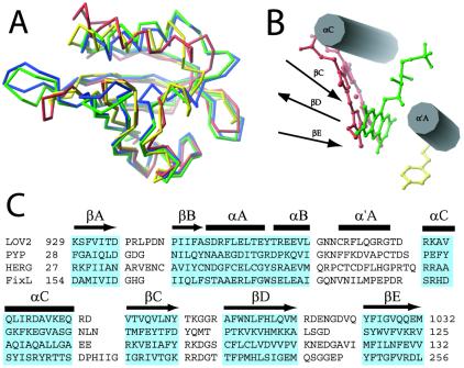
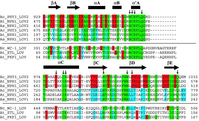
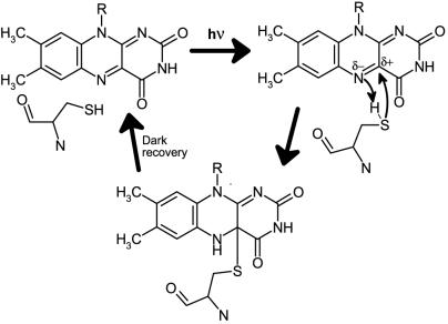
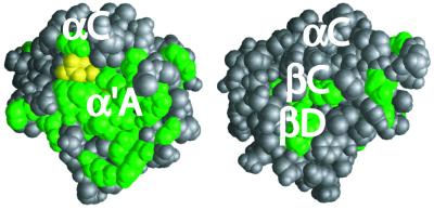
Phototropin, a major blue-light receptor for phototropism in seed plants, exhibits blue-light-dependent autophosphorylation and contains two light, oxygen, or voltage (LOV) domains and a serine/threonine kinase domain. The LOV domains share homology with the PER-ARNT-SIM (PAS) superfamily, a diverse group of sensor proteins. Each LOV domain noncovalently binds a single FMN molecule and exhibits reversible photochemistry in vitro when expressed separately or in tandem. We have determined the crystal structure of the LOV2 domain from the phototropin segment of the chimeric fern photoreceptor phy3 to 2.7-A resolution. The structure constitutes an FMN-binding fold that reveals how the flavin cofactor is embedded in the protein. The single LOV2 cysteine residue is located 4.2 A from flavin atom C(4a), consistent with a model in which absorption of blue light induces formation of a covalent cysteinyl-C(4a) adduct. Residues that interact with FMN in the phototropin segment of the chimeric fern photoreceptor (phy3) LOV2 are conserved in LOV domains from phototropin of other plant species and from three proteins involved in the regulation of circadian rhythms in Arabidopsis and Neurospora. This conservation suggests that these domains exhibit the same overall fold and share a common mechanism for flavin binding and light-induced signaling.


Figures

Figure 1
Figure 1
Adiantum phy3 domain and LOV2 structures. (A) Adiantum phy3 domain structure showing the N-terminal phytochrome chromophore domain bound to a phototropin. Residues forming the LOV2 construct are marked by arrows. (B) ribbon diagram of the phy3 LOV2 structure. The FMN cofactor is shown in the chromophore-binding pocket of LOV2 and is colored by elements: carbon, green; nitrogen, blue; oxygen, red; phosphorus, pink. C966 is at the N terminus of α′A and is colored yellow.
Figure 2
Figure 2
Chromophore of phy3 LOV2. (A) Simulated-annealing omit map of FMN from one of the four monomers in the asymmetric unit. The map is contoured at ±3.5σ (blue) and ±10σ (yellow), in which σ is the rms-deviation value of the electron density. Electron density distinguishes the dimethylbenzene and pyrimidine moieties of the isoalloaxazine ring and shows a +10σ feature over the terminal phosphate. (B) Stereo diagram of FMN–protein interactions. All residues and waters that hydrogen bond to or form van der Waals contact with FMN are shown. Hydrogen bonds are indicated by the dotted blue lines using a 2.6- to 3.5-Å range for hydrogen bonding. Atoms are colored as in Fig. 1 with the addition of sulfur as yellow, water molecules as light blue, and C(4a) of the isoalloxazine ring as pink.
Figure 3
Figure 3
Structural alignment of four PAS domains. (A) Least-squares superposition: FixL, red; PYP, yellow; HERG, blue; phy3 LOV2, green. (B) Comparison of positions of phy3 LOV2 (green), FixL (red), and PYP (yellow) chromophores within the chromophore-binding pocket of PAS domain. The secondary structural elements are represented schematically as cylinders and arrows. (C) Structure-based alignment of the sequences of these four PAS domains. Residues boxed in blue form secondary structural elements conserved among all four structures. These residues were used to optimize the least-squares fit shown in Fig. 3A. Secondary structure is noted above the alignment: β-strand, arrows; helix, bars.
Figure 4
Figure 4
Alignment of nine LOV-domain sequences and identification of flavin-interacting residues including LOV1 and LOV2 domains from Adiantum phy3 (GenBank accession no. BAA36192), Arabidopsis phototropin (GenBank accession no. AAC01753), oat phototropin (GenBank accession no. AAC05083), Arabidopsis ztl (GenBank accession no. AAF70288), Arabidopsis fkf1 (GenBank accession no. AAF32298), and Neurospora Wc-1 (GenBank accession no. Q01371). LOV2 residues that interact with FMN are marked with vertical arrows. Secondary structure of LOV2 is marked above alignment: β-strand, arrows; helix, bars. Green residues are conserved in all LOV1 and LOV2 domains included in the alignment as well as eight other LOV domains (not included because of space limitations) from rice phototropin (BAA84779), corn phototropin (T01353), rice phototropin-like protein nph1b (GenBank accession no. BAA84779) and Arabidopsis phototropin-like protein npl1 (GenBank accession no. AAC27293). Blue residues are conserved in all LOV1 domains, and red residues are conserved in all LOV2 domains from the above-listed proteins. This color scheme is applied to residues in Wc-1, ztl, and fkf1.
Figure 5
Figure 5
Proposed schematic mechanism for cysteine-C(4a) covalent adduct formation in response to light absorption by the LOV domain (see text).
Figure 6
Figure 6
Space-filling model of phy3 LOV2 showing the surface position of residues conserved in all LOV1 and LOV2 domains (green). Terminal phosphate of FMN is colored yellow. (Left) Model of the conserved face containing the 310-helix α′A. (Right) Model is rotated 180° about a vertical axis in the plane of Fig. 1.

References

    1. Singhal G S, Renger G, Sopory S K, Irrgang K-D, Govindjee . Concepts in Photobiology: Photosynthesis and Photomorphogenesis. Boston: Kluwer; 1999. pp. 775–929.
    1. Batschauer A. Planta. 1998;206:479–492. - PubMed
    1. Briggs WR, Huala E. Annu Rev Cell Dev Biol. 1999;15:33–62. - PubMed
    1. Fankhauser C, Chory J. Curr Biol. 1999;9:123–126. - PubMed
    1. Cashmore A R, Jarillo J A, Wu Y J, Liu D M. Science. 1999;284:760–765. - PubMed

Publication types

MeSH terms

Associated data

LinkOut - more resources