

Comparing protein-ligand interactions in solution and single crystals by Raman spectroscopy
- PMID: 11248022
- PMCID: PMC30597
- DOI: 10.1073/pnas.061029598
Comparing protein-ligand interactions in solution and single crystals by Raman spectroscopy
Abstract
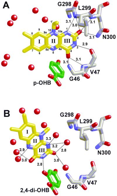
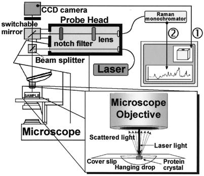
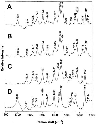
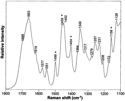
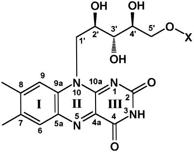
By using a Raman microscope, we show that it is possible to probe the conformational states in protein crystals and crystal fragments under growth conditions (in hanging drops). The flavin cofactor in the enzyme para-hydroxybenzoate hydroxylase can assume two conformations: buried in the protein matrix ("in") or essentially solvent-exposed ("out"). By using Raman difference spectroscopy, we previously have identified characteristic flavin marker bands for the in and out conformers in the solution phase. Now we show that the flavin Raman bands can be used to probe these conformational states in crystals, permitting a comparison between solution and crystal environments. The in or out marker bands are similar for the respective conformers in the crystal and in solution; however, significant differences do exist, showing that the environments for the flavin's isoalloxazine ring are not identical in the two phases. Moreover, the Raman-band widths of the flavin modes are narrower for both in and out conformers in the crystals, indicating that the flavin exists in a more limited range of closely related conformational states in the crystal than in solution. In general, the ability to compare detailed Raman data for complexes in crystals and solution provides a means of bridging crystallographic and solution studies.
Figures
References
-
- Palfey B A, Massey V. In: Comprehensive Biological Catalysis: A Mechanistic Reference. Sinnott M, editor. Vol. 4. San Diego: Academic; 1998.
-
- Palfey B A, Ballou D P, Massey V. In: Active Oxygen in Biochemistry. Valentine J S, Foote C S, Greenburg A, Lieberman J F, editors. Vol. 3. London: Blackie; 1995.
-
- Schreuder H A, Prick P A, Wierenga R K, Vriend G, Wilson K S, Hol W G, Drenth J. J Mol Biol. 1989;208:679–696. - PubMed
-
- Schreuder H A, van der Laan J M, Swarte M B, Kalk K H, Hol W G, Drenth J. Proteins. 1992;14:178–190. - PubMed
-
- Schreuder H A, Mattevi A, Obmolova G, Kalk K H, Hol W G, van der Bolt F J, van Berkel W J. Biochemistry. 1994;33:10161–10170. - PubMed
Publication types
MeSH terms
Substances
Grants and funding
LinkOut - more resources
Full Text Sources
