. 2001 Mar 13;98(6):3012-7.
doi: 10.1073/pnas.041619798. Epub 2001 Feb 27.

Solution structure of the antiapoptotic protein bcl-2

Affiliations

Solution structure of the antiapoptotic protein bcl-2

A M Petros et al. Proc Natl Acad Sci U S A. .

Abstract

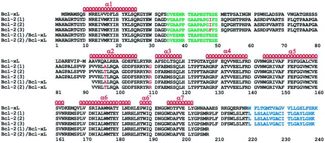
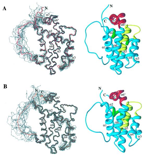
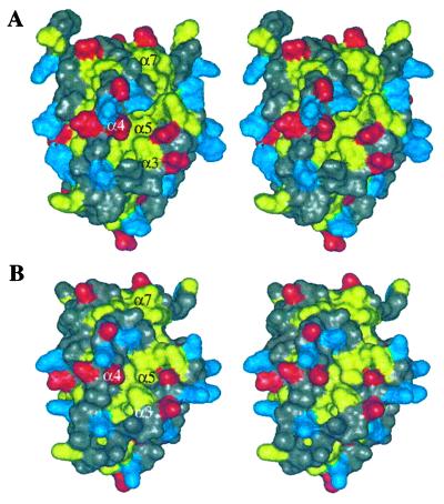
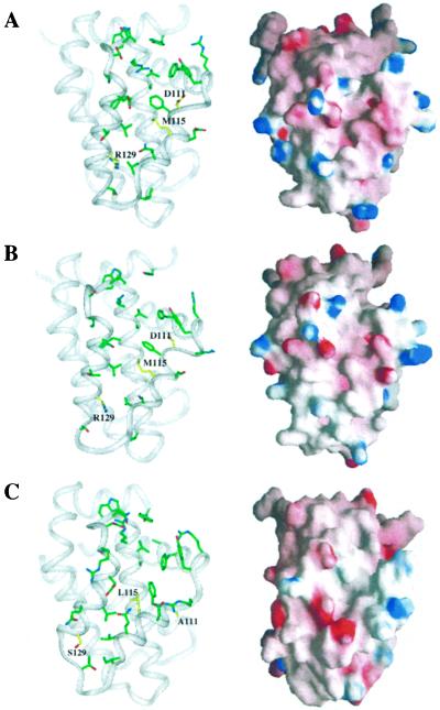
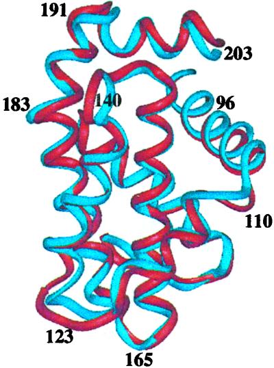
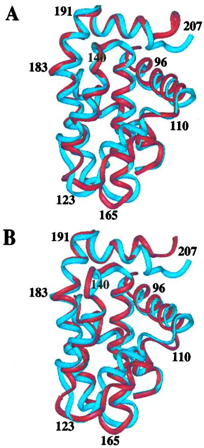
The structures of two isoforms of Bcl-2 that differ by two amino acids have been determined by NMR spectroscopy. Because wild-type Bcl-2 behaved poorly in solution, the structures were determined by using Bcl-2/Bcl-x(L) chimeras in which part of the putative unstructured loop of Bcl-2 was replaced with a shortened loop from Bcl-x(L). These chimeric proteins have a low pI compared with the wild-type protein and are soluble. The structures of the two Bcl-2 isoforms consist of 6 alpha-helices with a hydrophobic groove on the surface similar to that observed for the homologous protein, Bcl-x(L). Comparison of the Bcl-2 structures to that of Bcl-x(L) shows that although the overall fold is the same, there are differences in the structural topology and electrostatic potential of the binding groove. Although the structures of the two isoforms of Bcl-2 are virtually identical, differences were observed in the ability of the proteins to bind to a 25-residue peptide from the proapoptotic Bad protein and a 16-residue peptide from the proapoptotic Bak protein. These results suggest that there are subtle differences in the hydrophobic binding groove in Bcl-2 that may translate into differences in antiapoptotic activity for the two isoforms.


Figures

Figure 1
Figure 1
Sequence alignment of full-length Bcl-xL, the three isoforms of full-length Bcl-2 [denoted Bcl-2(1) (1,2), Bcl-2(2) (3,4), and Bcl-2(3) (5,6)], and the truncated Bcl-2/Bcl-xL chimeras used in this study. Amino acid differences between the Bcl-2 isoforms are shown in red, the truncated loop is shown in green, and the putative membrane-spanning region is shown in blue. α-helices previously identified in Bcl-xL are denoted above the sequence in red.
Figure 2
Figure 2
(A) Backbone (N, Cα, C′) superposition of 15 low-energy NMR-derived structures and Ribbons (47) depiction of the average minimized structure for Bcl-2(1). (B) Backbone (N, Cα, C′) superposition of 15 low-energy NMR-derived structures and Ribbons depiction of the average-minimized structure for Bcl-2(2). For the superpositions, the mean structure is shown in red. Helices are numbered with respect to those observed in the structure of Bcl-xL (20).
Figure 3
Figure 3
Solvent-accessible surface showing hydrophobic groove for Bcl-2(1) (A) and Bcl-2(2) (B). Leucine, isoleucine, valine, tyrosine, phenylalanine, and tryptophan residues are colored yellow, aspartate and glutamate are colored red, and lysine, arginine, and histidine are colored blue. All other residue types are colored gray.
Figure 4
Figure 4
Binding groove with key side chains and electrostatic [grasp (45)]) surface for Bcl-2(1) (A), Bcl-2(2) (B), and Bcl-xL (C). Residues that differ between the Bcl-2 proteins and Bcl-xL are highlighted in yellow.
Figure 5
Figure 5
Backbone superposition of Bcl-2(1) (red) with Bcl-2(2) (blue).
Figure 6
Figure 6
(A) Backbone superposition of Bcl-2(1) (red) with Bcl-xL (blue). (B) Backbone superposition of Bcl-2(2) (red) with Bcl-xL (blue).

References

    1. Tsujimoto Y, Finger L R, Yunis J, Nowell P C, Croce C M. Science. 1984;226:1097–1099. - PubMed
    1. Tsujimoto Y, Croce C M. Proc Natl Acad Sci USA. 1986;83:5214–5218. - PMC - PubMed
    1. Cleary M L, Sklar J. Proc Natl Acad Sci USA. 1985;82:7439–7443. - PMC - PubMed
    1. Cleary M L, Smith S D, Sklar J. Cell. 1986;47:19–28. - PubMed
    1. Bakhsi A, Jensen J P, Goldman P, Wright J J, McBride O W, Epstein A L, Korsmeyer S J. Cell. 1985;41:899–906. - PubMed

MeSH terms