. 2001 Mar 13;98(6):3086-91.
doi: 10.1073/pnas.061555698.

Herpes simplex virus DNA packaging sequences adopt novel structures that are specifically recognized by a component of the cleavage and packaging machinery

Affiliations

Herpes simplex virus DNA packaging sequences adopt novel structures that are specifically recognized by a component of the cleavage and packaging machinery

K Adelman et al. Proc Natl Acad Sci U S A. .

Abstract

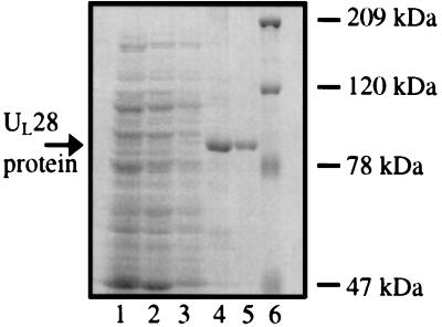
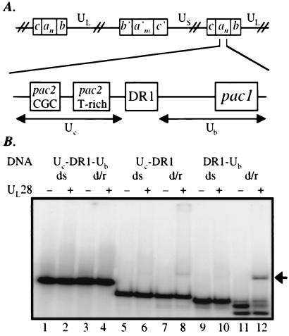
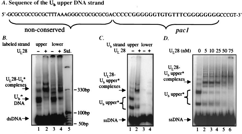
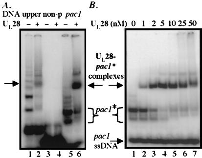
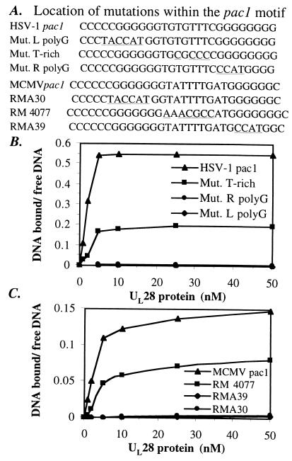
The product of the herpes simplex virus type 1 U(L)28 gene is essential for cleavage of concatemeric viral DNA into genome-length units and packaging of this DNA into viral procapsids. To address the role of U(L)28 in this process, purified U(L)28 protein was assayed for the ability to recognize conserved herpesvirus DNA packaging sequences. We report that DNA fragments containing the pac1 DNA packaging motif can be induced by heat treatment to adopt novel DNA conformations that migrate faster than the corresponding duplex in nondenaturing gels. Surprisingly, these novel DNA structures are high-affinity substrates for U(L)28 protein binding, whereas double-stranded DNA of identical sequence composition is not recognized by U(L)28 protein. We demonstrate that only one strand of the pac1 motif is responsible for the formation of novel DNA structures that are bound tightly and specifically by U(L)28 protein. To determine the relevance of the observed U(L)28 protein-pac1 interaction to the cleavage and packaging process, we have analyzed the binding affinity of U(L)28 protein for pac1 mutants previously shown to be deficient in cleavage and packaging in vivo. Each of the pac1 mutants exhibited a decrease in DNA binding by U(L)28 protein that correlated directly with the reported reduction in cleavage and packaging efficiency, thereby supporting a role for the U(L)28 protein-pac1 interaction in vivo. These data therefore suggest that the formation of novel DNA structures by the pac1 motif confers added specificity on recognition of DNA packaging sequences by the U(L)28-encoded component of the herpesvirus cleavage and packaging machinery.


Figures

Figure 1
Figure 1
Purification of the HSV-1 UL28 protein. Protein samples from each major step in the purification procedure were analyzed by SDS/PAGE. Lane 1, initial cell lysate; lane 2, supernatant after lysis; lane 3, supernatant from wash of the insoluble material; lane 4, Ni-nitrilotriacetic acid agarose beads, after incubation with the solubilized protein fraction and extensive washing; lane 5, eluate, after dialysis; lane 6, Bio-Rad high-molecular-weight protein marker.
Figure 2
Figure 2
(A) Diagram of the cis-acting sequences required for viral DNA cleavage/packaging. (Upper) One possible arrangement of an HSV-1 concatemer (as described in the introduction). (Lower) A detailed representation of the junction between two linear genomes and the sequences required for DNA cleavage/packaging. (B) EMSA analysis of the interaction between UL28 protein and DNA bearing pac sequences. DNA fragments spanning the regions Uc-DR1-Ub (lanes 1–4), Uc-DR1 (lanes 5–8), or DR1-Ub (lanes 9–12) were radiolabeled and either stored at −20°C [yielding the double-stranded (ds) probes present in lanes 1–2, 5–6, and 9–10] or boiled for 3 min and quickly transferred to ice before storage (probes labeled d/r, for denatured and reannealed; lanes 3–4, 7–8, and 11–12). The resultant DNA species were incubated at 5 nM in the absence or presence of UL28 protein (50 nM), and complexes were analyzed by EMSA. An arrow denotes the position at which the UL28 protein-DNA complexes migrate.
Figure 3
Figure 3
(A) The sequence of the Ub upper strand probe (63 nt), depicting components of nonconserved sequence as well as the conserved pac1 motif. (B) Analysis of UL28 protein binding to Ub DNA. DNA species formed by annealing complementary oligonucleotides corresponding to the Ub region were assayed for binding by UL28 protein. The labeled Ub upper strand was annealed to the unlabeled lower strand (lanes 1 and 2) or the labeled Ub lower strand annealed to an unlabeled upper strand (lanes 3 and 4). The DNA substrates (5 nM) were incubated in the presence or absence of 25 nM UL28 protein, and complexes were analyzed by EMSA. The relative mobility of the double-stranded DNA fragment (dsDNA), slowly migrating DNA species (Ub*), and specific complexes formed (UL28- Ub* complexes) are noted at the Left and can be compared with the DNA molecular size standard present in lane 5. (C) Investigation of strand specificity for the UL28 protein–Ub interaction. DNA species formed by the individual oligonucleotides corresponding to the upper (lanes 1 and 2) and lower (lanes 3 and 4) strands of the Ub region were assayed for interaction with UL28. The DNA probes (5 nM) were incubated in the absence or presence of the UL28 protein (10 nM), and complexes were analyzed EMSA. The slowly migrating species formed by the upper strand are labeled as Ub upper*. (D) Increasing concentrations of UL28 protein incubated with the Ub upper strand. A constant 5 nM Ub upper strand oligonucleotide DNA was incubated with UL28 at the final concentrations noted above each lane.
Figure 4
Figure 4
(A) Localization of the binding site for UL28 protein. Comparison was made between UL28 protein binding to oligonucleotides bearing the 63-nt Ub upper strand (lanes 1 and 2) and binding to probes comprising a 31-nt nonconserved sequence component (non-p, lanes 3 and 4) and the 27-nt pac1 motif (lanes 5 and 6). DNA present at 5 nM was incubated in the absence or presence of 10 nM UL28 protein and complexes analyzed by EMSA. The slowly migrating DNA species formed by the pac1 oligonucleotide are denoted as pac1*. (B) Determination of the binding affinity of UL28 protein for pac1* DNA. The pac1 oligonucleotide, present at 5 nM, was incubated with increasing concentrations of UL28 protein (as noted), and complexes were visualized by EMSA.
Figure 5
Figure 5
Correlation of in vitro binding affinity of UL28 protein with in vivo cleavage/packaging efficiency. (A) Sequence of the wild-type pac1 motif and mutant pac1 oligonucleotides. Nucleotide positions that differ from wild-type pac1 sequences are underlined. MCMV pac1 sequences are those that have been studied in vivo for their effects on cleavage/packaging (ref. and Michael McVoy, personal communication). The relative efficiency of cleavage/packaging observed for each mutant sequence as compared with MCMV wild-type pac1 (set at 100%) is as follows: RMA30, <2.5%; RM4077, 33%; RMA39, ≈3%. Binding affinity for each of the HSV-1 (B) and MCMV (C) pac1 oligonucleotides was determined. A concentration of 5 nM of each oligonucleotide was incubated with increasing concentrations of UL28 protein, and free DNA was separated from bound DNA by EMSA. The level of complex formation was determined by calculating the ratio of DNA bound to free DNA as a function of UL28 protein concentration (nM). Values shown represent the average of two to four experiments.

References

    1. Roizman B, Sears A E. In: Fundamental Virology. Fields B N, editor. New York: Raven; 1996. pp. 1048–1066.
    1. Spaete R R, Frenkel N. Cell. 1982;30:295–304. - PubMed
    1. Mocarski E S, Roizman B. Cell. 1982;31:89–97. - PubMed
    1. Stow N D, McMonagle E C, Davison A J. Nucleic Acids Res. 1983;11:8205–8220. - PMC - PubMed
    1. Varmuza S L, Smiley J R. Cell. 1985;41:793–802. - PubMed

Publication types

MeSH terms

LinkOut - more resources