. 2001 Mar 13;98(6):3092-7.
doi: 10.1073/pnas.041613598.

Reverse engineering the (beta/alpha )8 barrel fold

Affiliations

Reverse engineering the (beta/alpha )8 barrel fold

J A Silverman et al. Proc Natl Acad Sci U S A. .

Abstract

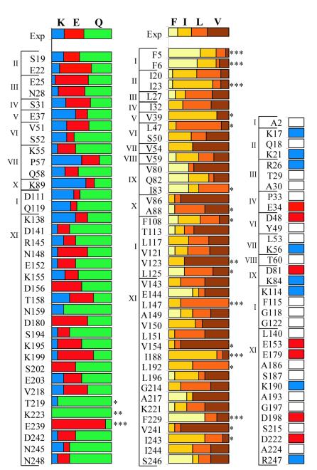
The (beta/alpha)(8) barrel is the most commonly occurring fold among protein catalysts. To lay a groundwork for engineering novel barrel proteins, we investigated the amino acid sequence restrictions at 182 structural positions of the prototypical (beta/alpha)(8) barrel enzyme triosephosphate isomerase. Using combinatorial mutagenesis and functional selection, we find that turn sequences, alpha-helix capping and stop motifs, and residues that pack the interface between beta-strands and alpha-helices are highly mutable. Conversely, any mutation of residues in the central core of the beta-barrel, beta-strand stop motifs, and a single buried salt bridge between amino acids R189 and D227 substantially reduces catalytic activity. Four positions are effectively immutable: conservative single substitutions at these four positions prevent the mutant protein from complementing a triosephosphate isomerase knockout in Escherichia coli. At 142 of the 182 positions, mutation to at least one amino acid of a seven-letter amino acid alphabet produces a triosephosphate isomerase with wild-type activity. Consequently, it seems likely that (beta/alpha)(8) barrel structures can be encoded with a subset of the 20 amino acids. Such simplification would greatly decrease the computational burden of (beta/alpha)(8) barrel design.


Figures

Figure 1
Figure 1
Phylogenetic conservation in TIM. The amino acid sequence of TIM is diagrammed according to secondary structure. The figure should be read from upper left to lower right. Secondary structure elements are designated at the left, with the following eight vertical columns corresponding to the eight β/α units of the protein. The yeast TIM sequence is shown in lowercase black letters (the first three amino acids are located at the lower right). The number of the first residue in each column is indicated at the top of the column. Conservation of a property in ≥75% of the sequences was required to assign a position to a conservation class. The green Hyd symbol indicates conservation of hydrophobic character [FILVAMGPWTYC] at that position. The cyan Pol symbol indicates conservation of polar character [HRKQNEDSTYC]. Red letters indicate conservation of a single amino acid or specific class of amino acids (symbols shown below). A blank space indicates no detectable conservation pattern. Conservation classes are defined as follows: Arom [FYW], Ncap [STDNQE], Beta [IVT], + [KRH], − [DE], Small [AG], 1mc [DN], 2mc [EQ], N-H [QNHW], φ,ψ [GP], StC [MRK], Mtl [HC], Nuc [STC].
Figure 2
Figure 2
Amino acid composition of functional sequences from a degenerate library. Amino acids observed in functional sequences from the degenerate library at phylogenetically polar positions (Left), phylogenetically hydrophobic positions (Center), and phylogenetically variable positions (Right). The brackets and roman numerals at the left of each graph indicate in which of the 11 libraries the position was mutated. The first row in each graph gives the average amino acid composition observed in the unselected library. Amino acids are color-coded as indicated at the top. Significant deviations from a random distribution (as determined by χ2 analysis) are indicated as follows: *, P < 5%; **, P < 1%; ***, P < 0.1%. Unconserved positions were mutated to a single amino acid. White bars indicate mutation to alanine, blue bars indicate mutation to lysine, and red bars indicate mutation to glutamate.
Figure 3
Figure 3
Structural environment of immutable residues. Top (A) and side (B) views of TIM showing the location of immutable residues. R189 is colored blue, D227 is colored red, and G209 and G228 are colored yellow. The backbone of the region corresponding to the C-terminal subdomain of tryptophan synthase is colored green. The N and C termini are indicated by black spheres. The active site is located toward the reader in A and at the top in B.

Comment in

  • Breaking open a protein barrel.
    Kallenbach N. Kallenbach N. Proc Natl Acad Sci U S A. 2001 Mar 13;98(6):2958-60. doi: 10.1073/pnas.071051798. Proc Natl Acad Sci U S A. 2001. PMID: 11248013 Free PMC article. Review. No abstract available.

References

    1. Reardon D, Farber G K. FASEB J. 1995;9:497–503. - PubMed
    1. Altamirano M M, Blackburn J M, Aguayo C, Fersht A R. Nature (London) 2000;403:617–622. - PubMed
    1. Jurgens C, Strom A, Wegener D, Hettwer S, Wilmanns M, Sterner R. Proc Natl Acad Sci USA. 2000;97:9925–9930. . (First Published August 15, 2000; 10.1073/pnas.160255397) - PMC - PubMed
    1. Kamtekar S, Schiffer J M, Xiong H, Babik J M, Hecht M H. Science. 1993;262:1680–1685. - PubMed
    1. Ponder J W, Richards F M. J Mol Biol. 1987;193:775–791. - PubMed

Publication types

Substances

LinkOut - more resources