

Toward a quantum-mechanical description of metal-assisted phosphoryl transfer in pyrophosphatase
- PMID: 11248042
- PMCID: PMC30617
- DOI: 10.1073/pnas.061612498
Toward a quantum-mechanical description of metal-assisted phosphoryl transfer in pyrophosphatase
Abstract
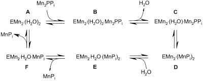
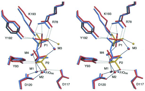
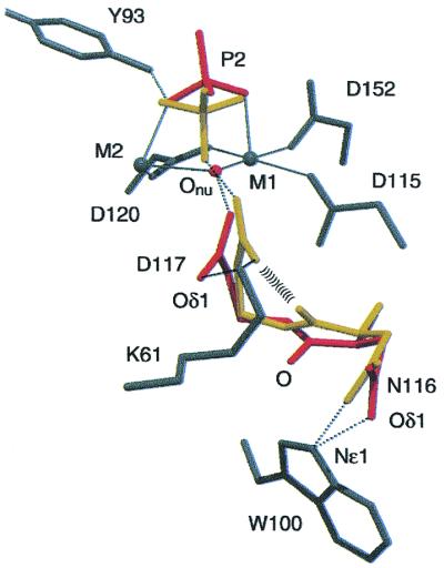
The wealth of kinetic and structural information makes inorganic pyrophosphatases (PPases) a good model system to study the details of enzymatic phosphoryl transfer. The enzyme accelerates metal-complexed phosphoryl transfer 10(10)-fold: but how? Our structures of the yeast PPase product complex at 1.15 A and fluoride-inhibited complex at 1.9 A visualize the active site in three different states: substrate-bound, immediate product bound, and relaxed product bound. These span the steps around chemical catalysis and provide strong evidence that a water molecule (O(nu)) directly attacks PPi with a pK(a) vastly lowered by coordination to two metal ions and D117. They also suggest that a low-barrier hydrogen bond (LBHB) forms between D117 and O(nu), in part because of steric crowding by W100 and N116. Direct visualization of the double bonds on the phosphates appears possible. The flexible side chains at the top of the active site absorb the motion involved in the reaction, which may help accelerate catalysis. Relaxation of the product allows a new nucleophile to be generated and creates symmetry in the elementary catalytic steps on the enzyme. We are thus moving closer to understanding phosphoryl transfer in PPases at the quantum mechanical level. Ultra-high resolution structures can thus tease out overlapping complexes and so are as relevant to discussion of enzyme mechanism as structures produced by time-resolved crystallography.
Figures


References
-
- Baykov A A, Cooperman B S, Goldman A, Lahti R. In: Progress in Molecular and Subcellular Biology. Schröder H C, Müller W E G, editors. Vol. 23. Heidelberg: Springer; 1999. pp. 127–150. - PubMed
-
- Belogurov G A, Fabrichniy I P, Pohjanjoki P, Kasho V N, Lehtihuhta E, Turkina M V, Cooperman B S, Goldman A, Baykov A A, Lahti R. Biochemistry. 2000;39:13931–13938. - PubMed
-
- Heikinheimo P, Lehtonen J, Baykov A, Lahti R, Cooperman B S, Goldman A. Structure. 1996;4:1491–1508. - PubMed
-
- Baykov A A, Fabrichniy I P, Pohjanjoki P, Zyryanov A B, Lahti R. Biochemistry. 2000;39:11939–11947. - PubMed
-
- Tuominen V, Heikinheimo P, Kajander T, Torkkel T, Hyytiä T, Kapyla J, Lahti R, Cooperman B S, Goldman A. J Mol Biol. 1998;284:1565–1580. - PubMed
Publication types
MeSH terms
Substances
Associated data
- Actions
- Actions
- Actions
- Actions
LinkOut - more resources
Full Text Sources
Other Literature Sources
Research Materials
