. 2001 Mar 13;98(6):3127-32.
doi: 10.1073/pnas.051633198. Epub 2001 Mar 6.

Structure of neurolysin reveals a deep channel that limits substrate access

Affiliations

Structure of neurolysin reveals a deep channel that limits substrate access

C K Brown et al. Proc Natl Acad Sci U S A. .

Abstract

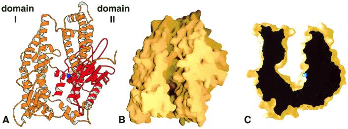
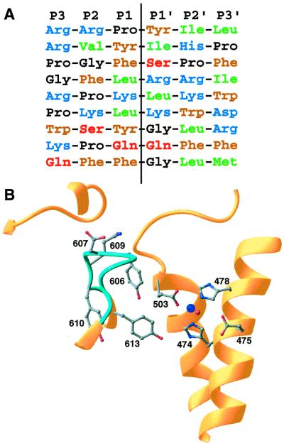
The zinc metallopeptidase neurolysin is shown by x-ray crystallography to have large structural elements erected over the active site region that allow substrate access only through a deep narrow channel. This architecture accounts for specialization of this neuropeptidase to small bioactive peptide substrates without bulky secondary and tertiary structures. In addition, modeling studies indicate that the length of a substrate N-terminal to the site of hydrolysis is restricted to approximately 10 residues by the limited size of the active site cavity. Some structural elements of neurolysin, including a five-stranded beta-sheet and the two active site helices, are conserved with other metallopeptidases. The connecting loop regions of these elements, however, are much extended in neurolysin, and they, together with other open coil elements, line the active site cavity. These potentially flexible elements may account for the ability of the enzyme to cleave a variety of sequences.


Figures

Figure 1
Figure 1
Overview of the rat neurolysin structure. (A) Ribbon view of the molecule looking down on the substrate-binding channel. The active site zinc ion is shown as a blue sphere, and the region structurally similar to other metallopeptidases is shown in red. (B) The same view shown as a molecular surface representation. (C) Cross-sectional view of the neurolysin substrate-binding channel. The depth of the channel at the active site (zinc ion in blue) is approximately 35 Å. This figure and all other ribbon and surface figures were prepared with molscript (50) and grasp (51).
Figure 2
Figure 2
Topology and the structurally conserved region. (A) Topology diagram of neurolysin with α-helices and β-strands numbered sequentially from the N terminus. Residue numbers for the beginning and end of larger secondary elements are given whenever possible. Dashed lines indicate points where regions have been separated for clarity. Portions shown in red are conserved in some other families of metallopeptidases. Ribbon diagrams for conserved regions of (B) neurolysin and (C) thermolysin also are shown. The active site zinc ion is in blue.
Figure 3
Figure 3
Model of substrate peptide binding. A molecular surface representation of neurolysin sectioned to show the large cavity at the bottom of the active-site channel is shown with the 13-residue substrate neurotensin modeled as described in the text. The N terminus of neurotensin is at the top.
Figure 4
Figure 4
Sequence selectivity. (A) Aligned sequences for known neurolysin cleavage sites. The sites listed are, top to bottom, from the neuropeptides neurotensin, angiotensin II, bradykinin, dynorphin A (residues 1–8), dynorphin A (residues 1–17), a second site in dynorphin A (residues 1–17), luteninizing hormone-releasing hormone, substance P, and a second site in substance P (8, 35). Residue types are indicated by different colors (blue = basic, brown = aromatic, green = aliphatic, black = proline or glycine, red = polar), and the hydrolysis position is indicated by the vertical line. (B) Details of the active site and nearby disordered loop (light blue; residues 600–612). The zinc cofactor is shown in dark blue and the catalytic water in red. Some side chains from residues in the mobile loop and active site are shown.

References

    1. Konkoy C S, Davis T P. Trends Pharmacol Sci. 1996;17:288–294. - PubMed
    1. Csuhai E, Safavi A, Thompson M W, Hersh L B. In: Proteolytic and Cellular Mechanisms in Prohormone and Neuropeptide Precursor Processing. Hook V, editor. Heidelberg: Springer; 1998. pp. 173–182.
    1. Checler F. In: Methods in Neurotransmitter and Neuropeptide Research. Nagatsu T, Parvez H, Naoi M, Parvez S, editors. Vol. 2. Amsterdam: Elsevier; 1993. pp. 375–418.
    1. Charli J L, Mendez M, Vargas M A, Cisneros M, Assai M, Joseph-Bravo P, Wilk S. Neuropeptides. 1989;14:191–196. - PubMed
    1. Vincent B, Dive V, Yiotakis A, Smadja C, Maldonado R, Vincent J-P, Checler F. Brit J Pharmacol. 1995;115:1053–1063. - PMC - PubMed

Publication types

Associated data