. 2001 Mar 13;98(6):3156-61.
doi: 10.1073/pnas.061032098.

p63 identifies keratinocyte stem cells

Affiliations

p63 identifies keratinocyte stem cells

G Pellegrini et al. Proc Natl Acad Sci U S A. .

Abstract

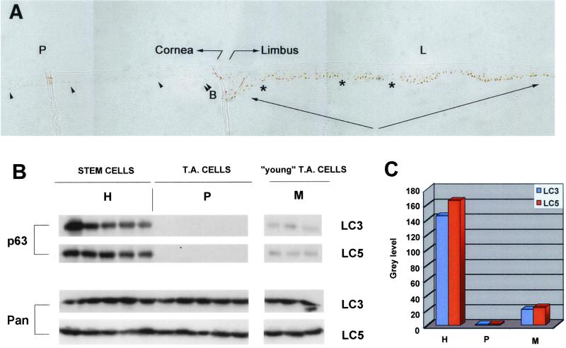
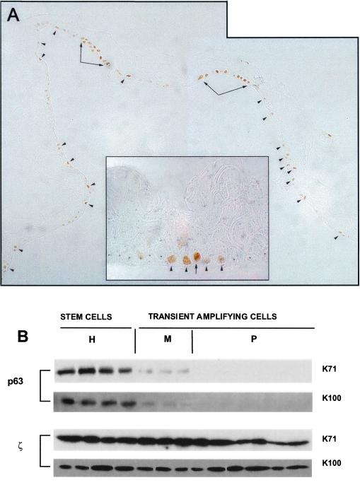
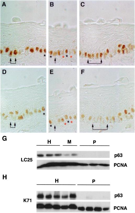
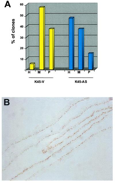
The proliferative compartment of stratified squamous epithelia consists of stem and transient amplifying (TA) keratinocytes. Some polypeptides are more abundant in putative epidermal stem cells than in TA cells, but no polypeptide confined to the stem cells has yet been identified. Here we show that the p63 transcription factor, a p53 homologue essential for regenerative proliferation in epithelial development, distinguishes human keratinocyte stem cells from their TA progeny. Within the cornea, nuclear p63 is expressed by the basal cells of the limbal epithelium, but not by TA cells covering the corneal surface. Human keratinocyte stem and TA cells when isolated in culture give rise to holoclones and paraclones, respectively. We show by clonal analysis that p63 is abundantly expressed by epidermal and limbal holoclones, but is undetectable in paraclones. TA keratinocytes, immediately after their withdrawal from the stem cell compartment (meroclones), have greatly reduced p63, even though they possess very appreciable proliferative capacity. Clonal evolution (i.e., generation of TA cells from precursor stem cells) is promoted by the sigma isoform of the 14-3-3 family of proteins. Keratinocytes whose 14-3-3final sigma has been down-regulated remain in the stem cell compartment and maintain p63 during serial cultivation. The identification of p63 as a keratinocyte stem cell marker will be of practical importance for the clinical application of epithelial cultures in cell therapy as well as for studies on epithelial tumorigenesis.


Figures

Figure 1
Figure 1
Expression of p63 by human corneal stem and TA cells. (A) Corneal biopsies spanning from the peripheral cornea (P) to the limbus (L) were stained with p63-specific mAb. Eight different inclusions from four different biopsies were analyzed. The transition from limbus to peripheral cornea was identified by the beginning of the Bowman's membrane (double arrowhead, B) and by immunostaining with Keratin-3-specific mAb (not shown). Note that the limbal basal layer (arrows) expresses p63, and that patches of p63 cells (asterisks) stagger p63+ cells. (B) Clonal analysis of subconfluent primary limbal cultures obtained from two different donors (LC3 and LC5). Cell extracts were prepared from cultures generated by holoclones (H), paraclones (P), and meroclones (M). Equal amounts of protein were fractionated on 7.5% SDS-polyacrylamide gels, transferred to PVDF filters, and immunostained with p63-specific mAb (p63) and with antibodies to pan-14-3-3 (Pan). Identical results were obtained with two other limbal strains (LC1 and LC25). (C) Densitometric analysis of the blots shown in B.
Figure 2
Figure 2
Expression of p63 by human epidermal stem and TA cells. (A) Cultured epidermal sheets prepared from primary keratinocytes were stained with p63-specific mAb. p63 was confined to the basal cell layer. Note that p63+ cells were located at intervals as single cells (arrowheads) or as patches (arrows), and were separated by stretches of p63 cells. At higher magnification (Inset), it is possible to observe cells expressing high levels of p63 (arrow) flanked with cells expressing lower levels of the protein (arrowheads). The latter cells are flanked by p63 cells (asterisks). (B) Clonal analysis of subconfluent primary epidermal cultures obtained from two different donors (K71 and K100). Cell extracts were prepared from cultures generated by holoclones (H), meroclones (M), and paraclones (P). Equal amounts of protein were fractionated on 7.5% SDS-polyacrylamide gels, transferred to PVDF filters and immunostained with p63-specific mAb (p63) and with polyclonal antibodies to 14-3-3ζ (ζ). p63 was high in holoclones, nearly absent in meroclones, and undetectable in paraclones; 14-3-3ζ was similar in all.
Figure 3
Figure 3
Expression of p63 and PCNA by keratinocyte stem and TA cells. (AF) Parallel sections of limbal biopsies were stained with p63-specific mAb (AC) and PCNA-specific mAb (DF). Arrows indicate basal cells that expressed p63, but not PCNA. Black asterisks indicate cells that expressed both p63 and PCNA. B and E note a p63+/PCNA+ cell (black asterisks) flanked by cells expressing PCNA, but not p63 (red asterisks). (G and H) Clonal analysis of subconfluent primary corneal (LC25) and epidermal (K71) cultures. Cell extracts were prepared from cultures generated by holoclones (H), meroclones (M), and paraclones (P). Equal amounts of protein were fractionated on 7.5% (G) or on 12.5% (H) SDS-polyacrylamide gels, transferred to PVDF filters and immunostained with p63-specific mAb (p63) and with PCNA-specific mAb (PCNA).
Figure 4
Figure 4
Expression of p63 in keratinocytes immortalized by transduction with antisense RNA for 14-3-3σ. The expression of 14-3-3σ was stably down-regulated in primary keratinocytes by transduction with defective retrovirus carrying a full-length human 14-3-3σ cDNA in antisense orientation (12). These cells maintained telomerase activity and became immortal (12). (A) Clonal analysis was performed on keratinocytes transduced with antisense σ DNA (K45-AS) or with an empty vector (K45-V). Note that the percentage of stem cells in the control K45-V cultures was 5% (H, yellow column), but rose to approximately 50% in K45-AS keratinocytes (H, blue column). (B) Cultured epidermal sheets prepared from antisense-σ-transduced keratinocytes were stained with p63-specific mAb. Note that most of basal cells express high levels of p63.

References

    1. Barrandon Y. Dev Biol. 1993;4:209–215.
    1. Fuchs E, Segre J A. Cell. 2000;100:143–155. - PubMed
    1. Watt F M, Hogan B L M. Science. 2000;287:1427–1430. - PubMed
    1. Lehrer M S, Sun T-T, Lavker R M. J Cell Sci. 1998;111:2867–2875. - PubMed
    1. Morris R J, Potten C S. Cell Prolif. 1994;27:279–289. - PubMed

Publication types

MeSH terms

Grants and funding