. 2001 Mar 13;98(6):3162-7.
doi: 10.1073/pnas.051494698.

Arp2/3 complex and actin dynamics are required for actin-based mitochondrial motility in yeast

Affiliations

Arp2/3 complex and actin dynamics are required for actin-based mitochondrial motility in yeast

I R Boldogh et al. Proc Natl Acad Sci U S A. .

Abstract

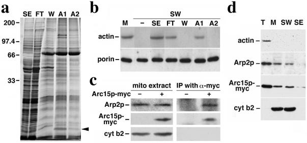
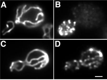
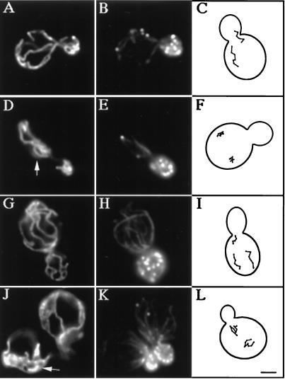
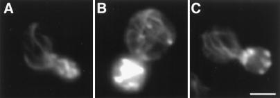
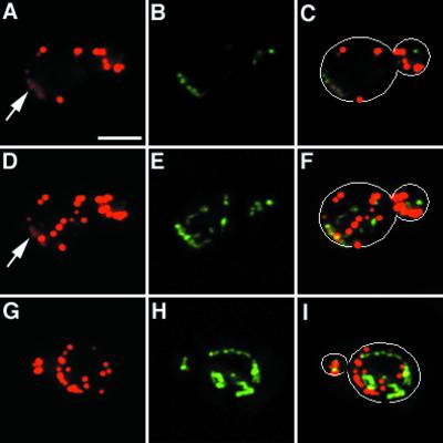
The Arp2/3 complex is implicated in actin polymerization-driven movement of Listeria monocytogenes. Here, we find that Arp2p and Arc15p, two subunits of this complex, show tight, actin-independent association with isolated yeast mitochondria. Arp2p colocalizes with mitochondria. Consistent with this result, we detect Arp2p-dependent formation of actin clouds around mitochondria in intact yeast. Cells bearing mutations in ARP2 or ARC15 genes show decreased velocities of mitochondrial movement, loss of all directed movement and defects in mitochondrial morphology. Finally, we observe a decrease in the velocity and extent of mitochondrial movement in yeast in which actin dynamics are reduced but actin cytoskeletal structure is intact. These results support the idea that the movement of mitochondria in yeast is actin polymerization driven and that this movement requires Arp2/3 complex.


Figures

Figure 1
Figure 1
Association of Arc15p and Arp2p subunits of Arp2/3 complex with mitochondria. (a) Silver-stained SDS gel of proteins recovered by actin affinity chromatography. SE, salt-extracted mitochondrial membrane proteins loaded onto the F-actin column; FT, column flow-through; W, proteins eluted with 200 mM KCl wash; A1, proteins eluted with 0.1 mM ATP; and A2, proteins eluted with 1 mM ATP. The sizes shown were determined by using molecular weight standards; the arrow points to the 15-kDa Arc15p-containing band in A1. (b) Restoration of mitochondrial actin-binding activity with column fractions. SE and column fractions (FT, W, A1, and A2) were added to salt-washed mitochondria (SW). Mitochondria were then separated from the mixture by centrifugation at 10,000 × g at 4°C. mABP activity in control mitochondria (M) and salt-washed mitochondria incubated with various column fractions was measured by using a sedimentation assay (16). Proteins recovered in the mitochondrial pellet were identified by Western blot analysis using antibodies raised against the mitochondrial marker, porin, and monoclonal antibody raised against actin (c4d6). (c) Immunoprecipitation of Arc15p-myc and Arp2p. Purified mitochondria were solubilized in the presence of 0.5% digitonin. The solubilized mitochondria were incubated with affinity-purified anti-myc antibody prebound to protein G-Sepharose beads. Immunoprecipitated proteins on the beads were identified by SDS/PAGE and Western blot analysis. Lanes 1–2, 25-μg digitonin-solubilized mitochondrial extracts from cells expressing untagged (−) or myc-tagged (+) Arc15p (DNY108 and DNY262). Lanes 3–4, immuno-precipitated proteins from 350-μg mitochondrial extracts from cells expressing untagged (−) or myc-tagged (+) Arc15p. (d) Actin-independent association of Arp2p and Arc15p with mitochondria. Isolated mitochondria from ARC15-MYC (DNY262) cells were washed with buffer containing 1 M KCl. Total cell lysate (T), mitochondria (M), salt washed mitochondria (SW), and salt extracted membrane proteins (SE) were evaluated for the levels of actin, Arp2p, Arc15p-myc, and cytochrome b2, a mitochondrial marker protein, by Western blot analysis. Proteins (30 μg) were loaded for T, M, and SW. SE was precipitated by adding trichloroacetic acid (TCA) to 10%. Precipitated proteins were resuspended to a volume equal to that of mitochondria (M). SE was loaded in the same volume as M.
Figure 2
Figure 2
Localization of Arp2p in yeast. DAUL1 cells expressing a fusion protein consisting of the mitochondrial signal sequence of citrate synthase 1 fused to GFP were grown to mid-log phase, fixed with paraformaldehyde alone (C and D), or with additional methanol/acetone (A and B), converted to spheroplasts, and stained for Arp2p by using an antibody raised against a conserved peptide sequence found in Arp2p, but not in actin or any other actin-related protein (30). (A and C) Mitochondria visualized by using CS1-GFP; (B and D) Arp2p visualized by indirect immunofluorescence. Bar = 1 μm.
Figure 3
Figure 3
Mitochondrial morphology and motility are abnormal in arc15-GFP and arp2-1 mutants. The ARP2 parent (AC), and arp2-1 mutant (DF) were grown to mid-log phase at 22°C and shifted to 37°C for 1 h. The ARC15 wild-type (GI) and arc15-GFP mutant cells (JL) were grown to mid-log phase at 30°C. Mitochondrial morphology (A, D, G, and J) and actin organization (B, E, H, and K) were determined in fixed cells by indirect immunofluorescence by using an antibody raised against mitochondrial outer membrane proteins, and rhodamine phalloidin. Arrowheads point to examples of abnormal mitochondrial morphology. (C, F, I, and L) Tracings of the pattern of mitochondrial movement in living cells stained with the membrane potential sensing dye, DiOC6 relative to the boundary of dividing yeast. Movements of mitochondria were followed by marking the tip of motile organelles during the time in which they remained in the plane of focus in time lapse images. The points denote the position of organelles at 20-sec intervals. Bar = 1 μm.
Figure 4
Figure 4
Actin organization in wild-type cells and stable actin mutants. Actin structure in wild-type ACT1 cells (A), act1-159 mutants (B), and act1-159 mutants treated with 15 μM Lat-A (C). Actin was stained with Alexa-phalloidin, and mitochondria were visualized by using CS1-GFP. Incubation with 15 μM Lat-A reduces the level of actin cables in act1-159 cells to that observed in wild-type ACT1 cells. Bar = 2 μm.
Figure 5
Figure 5
Actin cloud formation around mitochondria. The arp2-1 mutant and its wild-type parent expressing CS1-GFP were grown to mid-log phase at 22°C and treated with 0.1% sodium azide for 15 min. Cells were then fixed and stained with Alexa-phalloidin. Actin and mitochondria were visualized by optical sectioning, digital deconvolution, and three-dimensional reconstruction. A single optical section of a sodium azide-treated wild-type cell is shown in AC. Three-dimensional projection of the same cell is shown in DF. Three-dimensional projection of an arp2-1 mutant treated with sodium azide is shown in GI. (A, D and G) Alexa-phalloidin-labeled F-actin; (B, E, and H) GFP-labeled mitochondria; (C, F, and I) overlay of A and B, D and E, and G and H, respectively. Outlines of cells are superimposed over fluorescent images in C, F, and I. Arrows point to actin clouds surrounding mitochondria. Bar = 2 μm.

References

    1. Simon V R, Swayne T C, Pon L A. J Cell Biol. 1995;130:345–354. - PMC - PubMed
    1. Simon V R, Karmon S L, Pon L A. Cell Motil Cytoskeleton. 1997;37:199–210. - PubMed
    1. Yang H-C, Palazzo A, Swayne T C, Pon L A. Curr Biol. 1999;9:1111–1114. - PubMed
    1. Adams A E M, Pringle J. J Cell Biol. 1984;98:934–945. - PMC - PubMed
    1. Mulholland J, Preuss D, Moon A, Wong A, Drubin D, Botstein D. J Cell Biol. 1994;125:381–391. - PMC - PubMed

Publication types

MeSH terms