

Raft-partitioning of the ubiquitin ligases Cbl and Nedd4 upon IgE-triggered cell signaling
- PMID: 11248052
- PMCID: PMC30627
- DOI: 10.1073/pnas.051003498
Raft-partitioning of the ubiquitin ligases Cbl and Nedd4 upon IgE-triggered cell signaling
Abstract
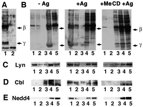
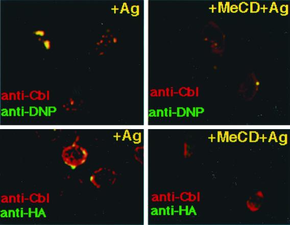
The high affinity receptor for IgE, FcepsilonRI on mast cells and basophils plays an essential role in immunological defense. Upon multivalent antigen binding, FcepsilonRI becomes phoshorylated by the protein-tyrosine kinase Lyn, as a result of receptor clustering in lipid rafts. FcepsilonRI has been shown to be ubiquitinated. Ubiquitination can lead to degradation by proteasomes, but it can also act as a sorting signal to internalize proteins destined to the endosomal/lysosomal pathway. We have analyzed whether FcepsilonRI ubiquitination takes place within rafts. We report biochemical and imaging evidence in rat basoleukemia cells for the presence of ubiquitinated FcepsilonRI in clustered rafts upon receptor activation. Moreover, we demonstrated that the ubiquitin ligases Cbl and Nedd4 colocalize with FcepsilonRI patches and showed that both ligases become associated with lipid rafts after activation of IgE signaling. Because Cbl is known to interact with the FcepsilonRI signaling complex, ubiquitination is likely to be an important parameter regulating IgE-triggered signaling occurring in rafts.
Figures


Similar articles
-
Negative regulation of FcepsilonRI-mediated mast cell activation by a ubiquitin-protein ligase Cbl-b.Blood. 2004 Mar 1;103(5):1779-86. doi: 10.1182/blood-2003-07-2260. Epub 2003 Nov 6. Blood. 2004. PMID: 14604964
-
Activation of Syk tyrosine kinase is required for c-Cbl-mediated ubiquitination of Fcepsilon RI and Syk in RBL cells.J Biol Chem. 2002 Oct 4;277(40):36940-7. doi: 10.1074/jbc.M204948200. Epub 2002 Jul 26. J Biol Chem. 2002. PMID: 12145291
-
Cbl family proteins: balancing FcεRI-mediated mast cell and basophil activation.Int Arch Allergy Immunol. 2011;156(1):16-26. doi: 10.1159/000322236. Epub 2011 Mar 29. Int Arch Allergy Immunol. 2011. PMID: 21447956 Review.
-
The adaptor molecule CIN85 regulates Syk tyrosine kinase level by activating the ubiquitin-proteasome degradation pathway.J Immunol. 2007 Aug 15;179(4):2089-96. doi: 10.4049/jimmunol.179.4.2089. J Immunol. 2007. PMID: 17675467
-
The Cbl family of ubiquitin ligases: critical negative regulators of tyrosine kinase signaling in the immune system.J Leukoc Biol. 2002 May;71(5):753-63. J Leukoc Biol. 2002. PMID: 11994499 Review.
Cited by
-
Mannheimia haemolytica leukotoxin binds to lipid rafts in bovine lymphoblastoid cells and is internalized in a dynamin-2- and clathrin-dependent manner.Infect Immun. 2007 Oct;75(10):4719-27. doi: 10.1128/IAI.00534-07. Epub 2007 Aug 6. Infect Immun. 2007. PMID: 17682044 Free PMC article.
-
Contrasting requirements for ubiquitylation during Fc receptor-mediated endocytosis and phagocytosis.EMBO J. 2002 Feb 1;21(3):251-8. doi: 10.1093/emboj/21.3.251. EMBO J. 2002. PMID: 11823418 Free PMC article.
-
Rhabdoviruses and the cellular ubiquitin-proteasome system: a budding interaction.J Virol. 2001 Nov;75(22):10623-9. doi: 10.1128/JVI.75.22.10623-10629.2001. J Virol. 2001. PMID: 11602704 Free PMC article.
-
The Role of the Host Ubiquitin System in Promoting Replication of Emergent Viruses.Viruses. 2021 Feb 26;13(3):369. doi: 10.3390/v13030369. Viruses. 2021. PMID: 33652634 Free PMC article. Review.
-
Lipid rafts in immune signalling: current progress and future perspective.Immunology. 2016 Sep;149(1):13-24. doi: 10.1111/imm.12617. Epub 2016 Jul 11. Immunology. 2016. PMID: 27153983 Free PMC article. Review.
References
Publication types
MeSH terms
Substances
LinkOut - more resources
Full Text Sources
Molecular Biology Databases
Miscellaneous
