. 2001 Mar 13;98(6):3220-5.
doi: 10.1073/pnas.051633398.

Cloning and mitochondrial localization of full-length D-AKAP2, a protein kinase A anchoring protein

Affiliations

Cloning and mitochondrial localization of full-length D-AKAP2, a protein kinase A anchoring protein

L Wang et al. Proc Natl Acad Sci U S A. .

Abstract

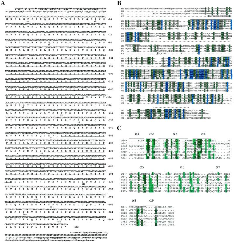
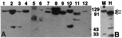
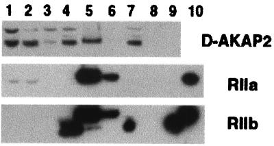
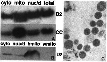
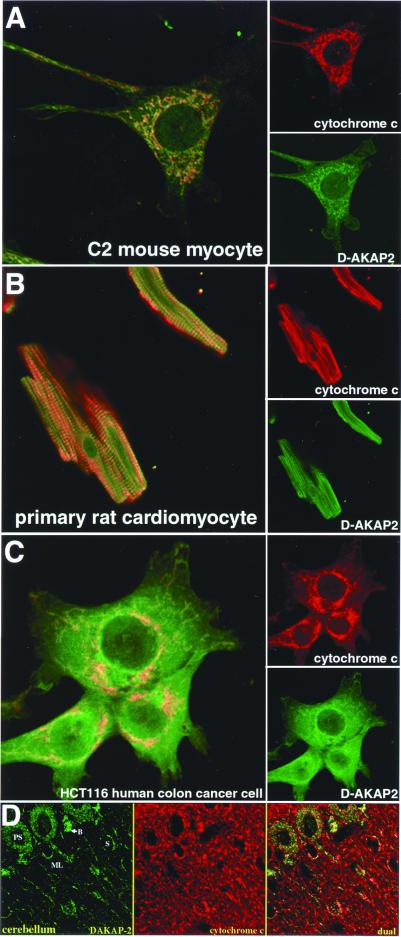
Differential compartmentalization of signaling molecules in cells and tissues is being recognized as an important mechanism for regulating the specificity of signal transduction pathways. A kinase anchoring proteins (AKAPs) direct the subcellular localization of protein kinase A (PKA) by binding to its regulatory (R) subunits. Dual specific AKAPs (D-AKAPs) interact with both RI and RII. A 372-residue fragment of mouse D-AKAP2 with a 40-residue C-terminal PKA binding region and a putative regulator of G protein signaling (RGS) domain was previously identified by means of a yeast two-hybrid screen. Here, we report the cloning of full-length human D-AKAP2 (662 residues) with an additional putative RGS domain, and the corresponding mouse protein less the first two exons (617 residues). Expression of D-AKAP2 was characterized by using mouse tissue extracts. Full-length D-AKAP2 from various tissues shows different molecular weights, possibly because of alternative splicing or posttranslational modifications. The cloned human gene product has a molecular weight similar to one of the prominent mouse proteins. In vivo association of D-AKAP2 with PKA in mouse brain was demonstrated by using cAMP agarose pull-down assay. Subcellular localization for endogenous mouse, rat, and human D-AKAP2 was determined by immunocytochemistry, immunohistochemistry, and tissue fractionation. D-AKAP2 from all three species is highly enriched in mitochondria. The mitochondrial localization and the presence of RGS domains in D-AKAP2 may have important implications for its function in PKA and G protein signal transduction.


Figures

Figure 1
Figure 1
(A) cDNA and deduced protein sequence of human D-AKAP2. Amino acid numbers are shown on the right. The two putative RGS domains are underlined. The star indicates a stop codon. Vertical arrows indicate the beginning of each numbered exon. Exons 1 and 15 extend beyond the cDNA sequence we have. (B) Comparison of the sequence of D-AKAP2 from four species: HU, human; MS, mouse; FL, fly, drosophila; CE, C. elegans. The PKA-binding region is indicated by stars (*). Horizontal lines above the sequence indicate the two putative RGS domains. (C) Alignment of the RGS domains of the N- and C-terminal portions of D-AKAP2. Comparison of the RGS domains of various RGS molecules in a multiple sequence alignment were generated by CLUSTAL W. The sequences include the N-terminal domain of D-AKAP2 (amino acids 125–242), C-terminal domain of D-AKAP2 (amino acids 379–502), rat RGS4 (amino acids 62–174, GenBank no. AF117211), human axin (amino acids 88–207, GenBank no. AF009674), P115 (amino acids 48–166, GenBank no. U64105), Drosophila Rho GEF2 (amino acids 930-1052, GenBank no. AF032870), and human GRK2 (amino acids 54–171, GenBank no. P25098). Also illustrated is the secondary structure of the largely α-helical RGS4, based on the x-ray crystal structure described by Tesmer et al. (24). α-Helical domains are represented by horizontal lines.
Figure 2
Figure 2
Expression of the D-AKAP2 protein in various mouse tissues. (A) Mouse tissue extracts were prepared as described in Experimental Procedures; 100 μg of total protein was loaded and probed with a polyclonal antibody against D-AKAP2 (anti-D-AKAP2). Antiserum, protein A, or antigen affinity-purified antibodies gave the same result, and none of the bands were detected by a number of unrelated control antibodies. The blots shown here used protein A purified antibody against the C terminus of mouse D-AKAP2. Lanes correspond to extracts from: white adipose tissue (1), BAT (2), skeletal muscle (3), tongue (4), small intestine (5), kidney (6), lung (7), brain (8), pancreas (9), heart (10), spleen (11), and liver (12). (B) Comparison of mouse and human full-length D-AKAP2. Human D-AKAP2 was in vitro translated by using a expression vector containing the cDNA as described in Experimental Procedures. It was then loaded onto SDS-PAGE along with a sample of mouse heart extract and probed with anti-D-AKAP2. M, mouse; H, human. The two arrows indicate the mobilities of the two full-length proteins. Lane 10 and M correspond to the same extract (*).
Figure 3
Figure 3
Isolation of complexes between D-AKAP2 and RII from mouse brain extract. cAMP pull-down assays were performed as described in Experimental Procedures. Lanes 1–3 are: total homogenate, supernatant, and resuspended pellet (a small fraction of the whole sample loaded). Lanes 4–6 and 7–9 are: flow through, cAMP elute and sample buffer elute after binding to the resin in the absence (lanes 4–6) or presence (lanes 7–9) of cAMP (entire amount of sample loaded). Lane 10 has purified RII standards.
Figure 4
Figure 4
Distribution of D-AKAP2 in mouse and rat adipose tissue fractions. Mitochondria were isolated from mouse BAT (A) or rat BAT (b) and WAT (w) (B) as described in Experimental Procedures to get the cytosolic supernatant, the nuclear and cell debris pellet and the mitochondria fraction. Then, 50 μg of total protein was loaded and probed with anti-D-AKAP2 (D2) or anti-cytochrome c (CC). Electron microscopy images were collected to indicate the quality of the isolated mitochondria and a representative image of BAT mitochondria is shown in C.
Figure 5
Figure 5
Immunocytochemistry and immunohistochemistry of D-AKAP2. (A) Mouse skeletal muscle cell line; (B) rat cardiomyocyte primary culture; (C) human colon cancer cell line; (D) cerebellum tissue. D-AKAP2 codistributes with cytochrome c in a subset of mitochondria. Strong codistribution is found in the Purkinje cell soma (PS), basket cells (B), and stellate cells (S) in the molecular layer (ML). Much weaker D-AKAP2 labeling is present in the dendrites and fibers of the molecular layer. Cells and tissue slices were stained with anti-D-AKAP2 (green) and anti-cytochrome c (red). Yellow areas indicate colocalization to the mitochondria.

References

    1. Cummings D E, Brandon E P, Planas J V, Motamed K, Idzerda R L, McKnight G S. Nature (London) 1996;382:622–626. - PubMed
    1. Jones P M, Sayed S B, Persaud S J, Burns C J, Gyles S, Whitehouse B J. J Mol Endocrinol. 2000;24:233–239. - PubMed
    1. Whitehouse B J, Abayasekara D R. J Mol Endocrinol. 1994;12:195–202. - PubMed
    1. Chen T C, Hinton D R, Zidovetzki R, Hofman F M. Lab Invest. 1998;78:165–174. - PubMed
    1. Tasken K, Skalhegg B S, Tasken K A, Solberg R, Knutsen H K, Levy F O, Sandberg M, Orstavik S, Larsen T, Johansen A K, et al. Adv Second Messenger Phosphoprotein Res. 1997;31:191–204. - PubMed

Publication types