. 2001 Mar 13;98(6):3288-93.
doi: 10.1073/pnas.041596598.

Ligand-independent assembly of recombinant human CD1 by using oxidative refolding chromatography

Affiliations

Ligand-independent assembly of recombinant human CD1 by using oxidative refolding chromatography

M M Altamirano et al. Proc Natl Acad Sci U S A. .

Abstract

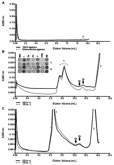
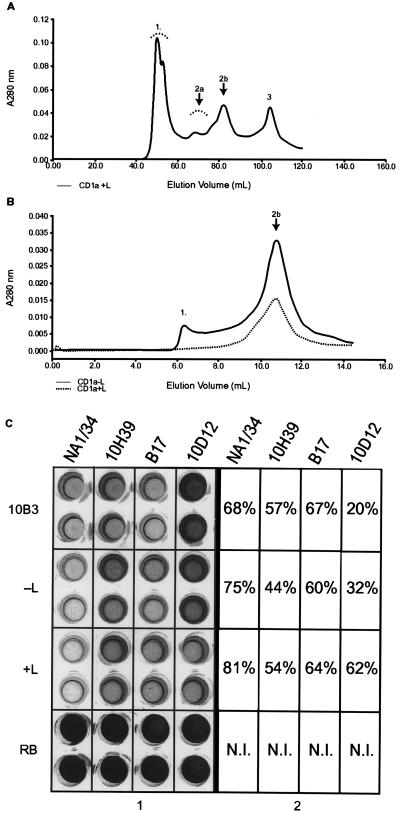
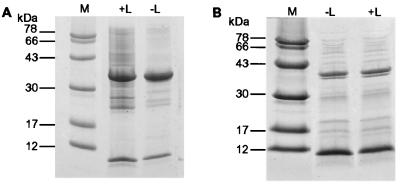
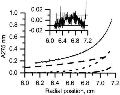
CD1 is an MHC class I-like antigen-presenting molecule consisting of a heavy chain and beta(2)-microglobulin light chain. The in vitro refolding of synthetic MHC class I molecules has always required the presence of ligand. We report here the use of a folding method using an immobilized chaperone fragment, a protein disulphide isomerase, and a peptidyl-prolyl cis-trans isomerase (oxidative refolding chromatography) for the fast and efficient assembly of ligand-free and ligand-associated CD1a and CD1b, starting with material synthesized in Escherichia coli. The results suggest that "empty" MHC class I-like molecules can assemble and remain stable at physiological temperatures in the absence of ligand. The use of oxidative refolding chromatography thus is extended to encompass complex multisubunit proteins and specifically to members of the extensive, functionally diverse and important immunoglobulin supergene family of proteins, including those for which a ligand has yet to be identified.


Figures

Figure 1
Figure 1
Refolding of CD1a under different conditions. (A) Elution profile from a Superdex-75 gel-filtration column of soluble supernatants when refolding was performed in the presence of either DsbA agarose (full line) or Ethanolamine agarose (broken line). The estimated molecular mass for peak 3 is 12 kDa. (B) Elution profile from an analytical Superdex-75 gel-filtration column of refolded CD1a supernatant by using oxidative refolding chromatography in the presence (full line) or absence (broken line) of exogenous ligand. The refolded CD1a (indicated by arrows as peaks 2a and 2b) corresponds to estimated molecular masses of ≈90 and 44 kDa, respectively. Peak 1 is a high molecular-mass aggregate. The estimated molecular mass for peak 3 is 12 kDa. (Inset) Antigenic activity of the unfractionated refolding mixture, MIX; peak 1 corresponding to aggregate, 1; peak 2a corresponding to dimeric CD1a, 2a; peak 2b corresponding to monomeric CD1a, 2b; and peak 3 corresponding to β2m, 3. The sCD1a-positive control and refolding-buffer-only negative control are labeled 10B3 and RB, respectively. +L refers to samples refolded in the presence of ligand and −L to those refolded without ligand. Each sample was tested in duplicate. Protein concentrations of the samples and positive control were approximately 1.0 mg⋅ml−1. (C) Elution profile from an analytical Superdex-75 gel-filtration column of CD1b supernatant refolded with (+L) and without (−L) specific ligand. Peaks (including 2a and 2b) correspond to molecular masses approximately similar to those above.
Figure 2
Figure 2
Optimization of the refolding procedure. (A) Scale-up of the CD1a refolding experiments and elution profile in a preparative Superdex-75 gel-filtration column. Estimated molecular masses of peaks correspond to those seen in the analytical gel-filtration column. (B) Rerun of peak 2b (obtained from scale-up, see Fig. 2A) in an analytical Superdex-75 column. (C) Antigenic characterization of peak 2b (obtained from fractionation of the CD1a refolding mixture supernatant). Samples were tested with four different conformation-sensitive CD1a mAbs: NA1/34, 10H39, B17, and 10D12. Numbers in the boxes refer to percentage of inhibition as compared with the refolding buffer-only (RB) negative control. The positive control is labeled 10B3. −L refers to samples refolded without specific ligand and +L to those refolded with ligand. The concentration of CD1a in the 10B3 positive control was approximately 1 mg⋅ml−1. N.I. indicates that no inhibition was seen in the negative control reference standard.
Figure 3
Figure 3
SDS/PAGE analysis of CD1a and -b refolding mixtures. (A) 15% SDS/PAGE analysis of CD1a refolding mixtures carried out with (+L) and without (−L) synthetic ligand, demonstrating bands of the expected molecular massas for the CD1a heavy and β2m light chain. Molecular mass marker standards are as indicated. (B) As above, but for CD1b.
Figure 4
Figure 4
Equilibrium sedimentation of refolded CD1a. Experiments were performed at 10,000 rpm, 4°C, by using 50 mM Tris/60 mM GuHCl/2% glycerol/1 mM EDTA. Data are for the CD1a −L fraction. Data for the +L complex were very similar. Data were fitted to a model-free equation that assumes that the CD1a complex can oligomerize, and that there is an excess of β2m. Heavy chain alone was not seen, indicating that the CD1a/β2m association is very tight, or/and that nonassociated heavy chain is insoluble. Logarithms of oligomerization constants (for dimer formation) were estimated to be 4.63 ± 0.1 for empty (−L) and 5.07 ± 0.1 for ligand-loaded (+L) complexes (Kd = 23 and 8.5 μM, respectively). Black line, experimental data and theoretical fit; dashed line, β2m alone; dotted line, CD1a; dashed-dotted line, CD1a dimer. The baseline contribution (0.064) is not shown. (Inset) Residues (Aexp-Afit) of the fit (A275 units). The horizontal lines at ± 0.01 represent the typical limits of stability in the instrument baseline.
Figure 5
Figure 5
C/D spectra (260–210 nm) of refolded/purified CD1a. Spectra were recorded in 25 mM potassium phosphate buffer (pH 8.0) containing 60 mM GuHCl and 2% glycerol. Black circles, β2m alone (0.15 mg⋅ml−1); crosses, CD1a (−L) (0.15 mg⋅ml−1); white triangles, CD1a (+L) (0.30 mg⋅ml−1); black triangles, CD1a dimer (−L) (0.35 mg⋅ml−1).

Comment in

Similar articles

Cited by

References

    1. Calabi, F. and Milstein, C. (2001) Semin. Immunol., in press. - PubMed
    1. Porcelli S A, Modlin R L. Annu Rev Immunol. 1999;17:297–329. - PubMed
    1. Calabi F, Yung Yu C, Bilsland C A G, Milstein C. In: CD1: From Structure to Function. Immunogenetics of the Major Histocompatibility Complex. Srivastava R, Ram B, Tyle P, editors. NY: VCH; 1991.
    1. Burdin N, Kronenberg M. Curr Opin Immunol. 1999;3:326–331. - PubMed
    1. Balk S P, Burke S, Polischuk J E, Frantz M E, Yang L, Porcelli S, Colgan S P, Blumberg R S. Science. 1994;265:259–262. - PubMed

Publication types