. 2001 Mar 13;98(6):3294-8.
doi: 10.1073/pnas.051604498.

Human CD1d-glycolipid tetramers generated by in vitro oxidative refolding chromatography

Affiliations

Human CD1d-glycolipid tetramers generated by in vitro oxidative refolding chromatography

A Karadimitris et al. Proc Natl Acad Sci U S A. .

Abstract

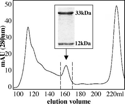
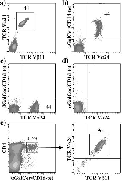
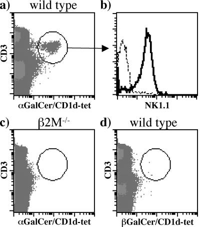
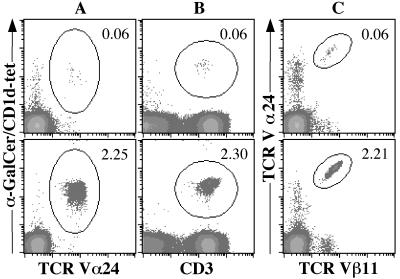
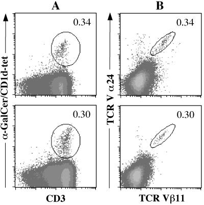
CD1 molecules are specialized in presenting lipids to T lymphocytes, but identification and isolation of CD1-restricted lipid specific T cells has been hampered by the lack of reliable and sensitive techniques. We here report the construction of CD1d-glycolipid tetramers from fully denatured human CD1d molecules by using the technique of oxidative refolding chromatography. We demonstrate that chaperone- and foldase-assisted refolding of denatured CD1d molecules and beta(2)-microglobulin in the presence of synthetic lipids is a rapid method for the generation of functional and specific CD1d tetramers, which unlike previously published protocols ensures isolation of CD1d tetramers loaded with a single lipid species. The use of human CD1d-alpha-galactosylceramide tetramers for ex vivo staining of peripheral blood lymphocytes and intrahepatic T cells from patients with viral liver cirrhosis allowed for the first time simultaneous analysis of frequency and specificity of natural killer T cells in human clinical samples. Application of this protocol to other members of the CD1 family will provide powerful tools to investigate lipid-specific T cell immune responses in health and in disease.


Figures

Figure 1
Figure 1
Purification of refolded CD1d–β2m monomers loaded with α-GalCer. FPLC and UV-light absorption profile at 280 nm wave length of a refolding mix containing CD1d and β2m in the presence of α-GalCer. The arrow indicates the CD1d–β2m monomeric complexes. (Inset) SDS/PAGE analysis of proteins eluted between 155 and 170 ml, which demonstrates the presence of two proteins of 33 and 12 kDa, corresponding to unglycosylated CD1d and β2m, respectively.
Figure 2
Figure 2
Specificity of CD1d–α-GalCer tetramers. (ad) Vα24+/Vβ11+ T cells were expanded after stimulation of PBMC with α-GalCer and were stained with CD1d tetramers and antibodies shown in the figure. Cells in d were incubated first with unlabeled anti-Vα24 and anti-Vβ11 antibodies and subsequently stained with FITC-conjugated anti-Vα24 antibody and CD1d–α-GalCer tetramers. All density plots were gated on the CD3+ propidium iodide cells. (e) CD4+ CD1d–α-GalCer tetramer+ cells were sorted from an in vitro-expanded α-GalCer-stimulated T cell line containing 0.59% positive cells. Enrichment for NKT cells was confirmed by using anti-Vα24 and -Vβ11 antibodies after two rounds of stimulation.
Figure 3
Figure 3
Staining of mouse NK1.1 cells by human CD1d–α-GalCer tetramers. (a and c) Splenocytes from wild-type C57BL/6 mice and β2m−/− mice were stained with human CD1d–α-GalCer tetramers and antibodies specific to CD3 and NK1.1. (b) Cells in histogram plot were gated on the CD3+ CD1d–α-GalCer+ tetramer+ cell population shown in a. Dashed line shows staining with a control antibody, and the continuous line shows staining with anti-NK1.1 (d) As a negative control, splenocytes from wild-type C57BL/6 mice were stained with CD1d–β-GalCer tetramers and anti-CD3 antibody.
Figure 4
Figure 4
Detection of CD1d–α-GalCer-specific T cells in peripheral blood from patients with HCV. Aliquots from each blood sample were stained with CD1d–α-GalCer tetramer and anti-Vα24 antibody (A), CD1d–α-GalCer tetramer and anti-CD3 antibody (B), or anti-Vα24 and -Vβ11 antibodies (C). Percentages of double-positive cells (circled regions) are indicated on each density plot. Top row corresponds to patient 1, and the bottom row corresponds to patient 5.
Figure 5
Figure 5
Staining of intrahepatic lymphocytes in patients with HCV and HBV infection. Samples from patients (P9, top row; P11, bottom row) were stained with CD1d–α-GalCer tetramers and anti-CD3 antibody (A) or anti-Vα24 and -Vβ11 antibodies (B). Percentages of CD1d–α-GalCer tetramer+/CD3+ and Vα24+/Vβ11+ cells are shown.

Comment in

Similar articles

Cited by

References

    1. Porcelli S A, Modlin R L. Annu Rev Immunol. 1999;17:297–329. - PubMed
    1. Bilsland C A, Milstein C. Eur J Immunol. 1991;21:71–78. - PubMed
    1. Calabi F, Jarvis J M, Martin L, Milstein C. Eur J Immunol. 1989;19:285–292. - PubMed
    1. Albertson D G, Fishpool R, Sherrington P, Nacheva E, Milstein C. EMBO J. 1988;7:2801–2805. - PMC - PubMed
    1. Martin L H, Calabi F, Milstein C. Proc Natl Acad Sci USA. 1986;83:9154–9158. - PMC - PubMed

Publication types