. 2001 Mar 13;98(6):3328-33.
doi: 10.1073/pnas.041614798.

Myocyte-enriched calcineurin-interacting protein, MCIP1, inhibits cardiac hypertrophy in vivo

Affiliations

Myocyte-enriched calcineurin-interacting protein, MCIP1, inhibits cardiac hypertrophy in vivo

B A Rothermel et al. Proc Natl Acad Sci U S A. .

Abstract

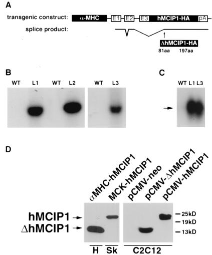
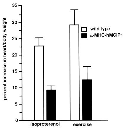
Signaling events controlled by calcineurin promote cardiac hypertrophy, but the degree to which such pathways are required to transduce the effects of various hypertrophic stimuli remains uncertain. In particular, the administration of immunosuppressive drugs that inhibit calcineurin has inconsistent effects in blocking cardiac hypertrophy in various animal models. As an alternative approach to inhibiting calcineurin in the hearts of intact animals, transgenic mice were engineered to overexpress a human cDNA encoding the calcineurin-binding protein, myocyte-enriched calcineurin-interacting protein-1 (hMCIP1) under control of the cardiac-specific, alpha-myosin heavy chain promoter (alpha-MHC). In unstressed mice, forced expression of hMCIP1 resulted in a 5-10% decline in cardiac mass relative to wild-type littermates, but otherwise produced no apparent structural or functional abnormalities. However, cardiac-specific expression of hMCIP1 inhibited cardiac hypertrophy, reinduction of fetal gene expression, and progression to dilated cardiomyopathy that otherwise result from expression of a constitutively active form of calcineurin. Expression of the hMCIP1 transgene also inhibited hypertrophic responses to beta-adrenergic receptor stimulation or exercise training. These results demonstrate that levels of hMCIP1 producing no apparent deleterious effects in cells of the normal heart are sufficient to inhibit several forms of cardiac hypertrophy, and suggest an important role for calcineurin signaling in diverse forms of cardiac hypertrophy. The future development of measures to increase expression or activity of MCIP proteins selectively within the heart may have clinical value for prevention of heart failure.


Figures

Figure 1
Figure 1
Design and expression of the α−MHC-hMCIP1 transgene. (A) Schematic illustration of components of the transgene, including a 5.5-kb α−MHC promoter fragment with three noncoding exons (E1, E2, and E3) and intervening nontranscribed segments of the α−MHC gene, followed by a full-length human MCIP1 cDNA with a carboxyl terminal epitope tag (HA) and a polyadenylation (pA) signal from the human GH gene. The lower line illustrates the unexpected pattern of mRNA splicing observed in vivo, resulting in translation of a truncated protein (ΔhMICP1) initiated at amino acid 81 relative to the wild-type (WT) protein. (B) Southern blot of genomic DNA from WT mice and each of three lines (L1, L2, and L3) of α−MHC-hMCIP1 transgenic mice by using a probe specific to the human MCIP1 gene sequence. (C) Northern blot of heart mRNA from WT and two of the α−MHC-hMCIP1 transgenic mice lines (L1 and L3) by using a probe specific to human MCIP1 mRNA. Transgene expression was higher in the L2 line (data not shown). The arrow indicates the anomalously spliced 1.2-kb α−MHC-hMCIP1 transgene transcript (ΔhMCIP). Expression of the transgene was at least an order of magnitude higher than the endogenous 2.2-kb mMCIP1 transcript (data not shown). (D) Western blots probed to detect the hMCIP-HA protein in extracts from an L1 α−MHC-hMCIP1 transgenic heart (H) showing the 14-kDa truncated ΔhMCIP1 protein product. A positive control for comparison to a correctly spliced full length 24-kDa hMCIP1 protein was drawn from a skeletal muscle extract (Sk) from a line of transgenic mice expressing hMCIP1 under the control of the muscle creatine kinase promoter (MCK-hMCIP1). C2C12 cells were transfected with plasmids expressing the indicated products for size comparisons [pCMV-neo, pCMV-ΔhMCIP1 (amino acids 81 to 197) and pCMV-hMCIP1 (amino acids 1 to 197)].
Figure 2
Figure 2
Functional activity of the truncated ΔhMCIP1 protein product compared with full-length hMCIP1, assessed by inhibition of calcineurin-dependent activation in C2C12 myoblasts. C2C12 cells were transfected with a luciferase reporter gene driven by a calcineurin-responsive IL-2 promoter (IL-2-Luc). Cells were cotransfected with a constitutively active calcineurin expression plasmid (pCMV-CnA*) and either an empty control vector (pCMV-neo) or the indicated MCIP-encoding plasmid (pCMV-hMCIP1 or pCMV-ΔhMCIP1).
Figure 3
Figure 3
Effects of hMCIP1 on cardiac hypertrophy induced by activated calcineurin. (A) Gross morphology of hearts from wild-type, α−MHC-hMCIP1 transgenic, α−MHC-CnA* transgenic, and doubly transgenic (α−MHC-hMCIP1 × α−MHC-CnA*) mice aged 20 weeks. (B) M-mode echocardiographic analysis from male littermates of the indicated genotypes at an age of 20 weeks. Heart rate was markedly slowed in the α-MHC-CnA* animals relative to mice of other genotypes. Echocardiographic measurements of these animals are found in Table 2. (C) RNA dot blot analysis of gene expression of GAPDH, α-MHC, β-MHC, α-skeletal actin, and ANF in hearts of mice of the indicated genotypes.
Figure 4
Figure 4
Effects of hMCIP1 on cardiac hypertrophy in response to β-adrenergic receptor stimulation or exercise in intact animals. The graph indicates the percentage increase in htw/bw of male wild-type (n = 7) and α−MHC-hMCIP1 transgenic (n = 9) mice subjected to chronic infusion of isoproterenol over 7 days or the percentage increase in htw/bw of male wild-type (n = 4) and α−MHC-hMCIP1 transgenic (n = 4) mice after 28 days of voluntary wheel running exercise. The α−MHC-hMCIP1 mice developed significantly less cardiac hypertrophy in response to either isoproterenol or exercise (P < 0.05).

Comment in

References

    1. Clipstone N A, Crabtree G R. Nature (London) 1992;357:695–697. - PubMed
    1. Friday B B, Horsley V, Pavlath G K. J Cell Biol. 2000;149:657–666. - PMC - PubMed
    1. Chin E R, Olson E N, Richardson J A, Yang Q, Humphries C, Shelton J M, Wu H, Zhu W, Bassel-Duby R, Williams R S. Genes Dev. 1998;12:2499–2509. - PMC - PubMed
    1. Wu H, Naya F J, McKinsey T A, Mercer B, Shelton J M, Chin E R, Simard A R, Michel R N, Bassel-Duby R, Olson E N, Williams R S. EMBO J. 2000;19:1963–1973. - PMC - PubMed
    1. Musaro A, McCullagh K J, Naya F J, Olson E N, Rosenthal N. Nature (London) 1999;400:581–585. - PubMed

Publication types

MeSH terms

LinkOut - more resources