. 2001 Mar 13;98(6):3352-7.
doi: 10.1073/pnas.061615598.

Heparin and cancer revisited: mechanistic connections involving platelets, P-selectin, carcinoma mucins, and tumor metastasis

Affiliations

Heparin and cancer revisited: mechanistic connections involving platelets, P-selectin, carcinoma mucins, and tumor metastasis

L Borsig et al. Proc Natl Acad Sci U S A. .

Abstract

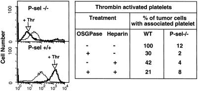
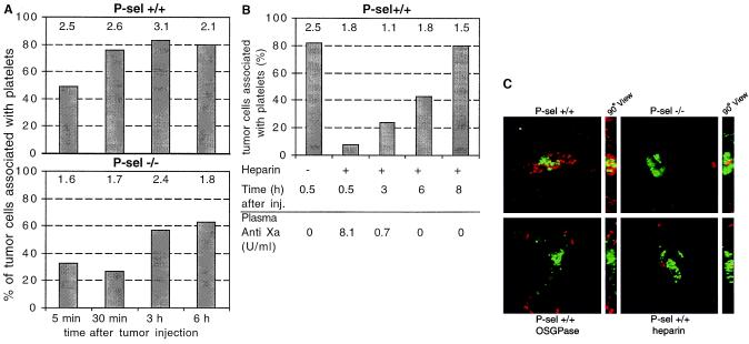
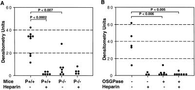
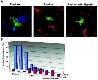
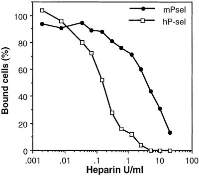
Independent studies indicate that expression of sialylated fucosylated mucins by human carcinomas portends a poor prognosis because of enhanced metastatic spread of tumor cells, that carcinoma metastasis in mice is facilitated by formation of tumor cell complexes with blood platelets, and that metastasis can be attenuated by a background of P-selectin deficiency or by treatment with heparin. The effects of heparin are not primarily due to its anticoagulant action. Other explanations have been suggested but not proven. Here, we bring together all these unexplained and seemingly disparate observations, showing that heparin treatment attenuates tumor metastasis in mice by inhibiting P-selectin-mediated interactions of platelets with carcinoma cell-surface mucin ligands. Selective removal of tumor mucin P-selectin ligands, a single heparin dose, or a background of P-selectin deficiency each reduces tumor cell-platelet interactions in vitro and in vivo. Although each of these maneuvers reduced the in vivo interactions for only a few hours, all markedly reduce long-term organ colonization by tumor cells. Three-dimensional reconstructions by using volume-rendering software show that each situation interferes with formation of the platelet "cloak" around tumor cells while permitting an increased interaction of monocytes (macrophage precursors) with the malignant cells. Finally, we show that human P-selectin is even more sensitive to heparin than mouse P-selectin, giving significant inhibition at concentrations that are in the clinically acceptable range. We suggest that heparin therapy for metastasis prevention in humans be revisited, with these mechanistic paradigms in mind.


Figures

Figure 1
Figure 1
Effects of heparin and OSGPase treatment on P-selectin-mediated tumor–platelet interactions in vitro. Mouse platelets were isolated from citrated blood as described (44) and calcein-AM labeled. Tumor cells were detached with PBS/2 mM EDTA, washed in HBSS (Sigma), and 200,000 cells mixed in HBSS with labeled mouse platelets (5 × 10⁷) with/without heparin (20 units/ml final) and incubated for 5 min. Tumor cells were analyzed by flow cytometry, to quantitate attachment of calcein-labeled platelets with or without preactivation with mouse thrombin (marked as Thr, 0.2 units/ml added 3 min before flow cytometry analysis). Tumor cells were also studied with or without prior mucin removal by OSGPase (see Materials and Methods). Summarized data are presented regarding the effects of heparin, OSGPase treatment, or a combination. The apparent reduction of P-selectin negative platelet interactions after thrombin is actually within the range of variation of negative results for this experiment, which was repeated several times.
Figure 2
Figure 2
Effects of heparin, OSGPase treatment, and P-selectin deficiency on tumor cell–platelet interactions in vivo. Mice were intravenously injected with calcein AM-labeled LS180 cells (green fluorescence) and killed at different time points. Frozen sections of the lungs were stained with biotinylated anti-CD41 Ab for platelets (Cy3 streptavidin for red fluorescence) as described in Materials and Methods. Platelet association with tumor cells was quantitated by evaluation of sections by using conventional epifluorescence microscopy (×40 objective, evaluating 16 random fields) (A) Time dependency of tumor–platelet complex formation in P-selectin+/+ and P-selectin−/− mice. (B) Heparin effect on tumor–platelet complex formation in P-sel+/+ mice 100 units of heparin was injected intravenously 30 min before the LS180 cells. In each case, numbers above the bars represent the mean number of tumor cells seen per high-power field. Heparin in circulation was measured by an anti-Xa assay of plasma (data below the graph, kindly performed by Dzung Le, University of California, San Diego). (C) Examples from deconvolutional microscopy of typical tumor–platelet complexes observed in lung sections 30 min after LS180 injection. OSGPase indicates tumor cells treated with OSGPase before injection. Heparin indicates heparin injection 30 min before tumor cell injection.
Figure 3
Figure 3
Effects of Heparin, OSGPase treatment, and P-selectin deficiency on the establishment and growth of tumor metastasis. Mice were intravenously injected with 3–4 × 10⁵ tumor cells and killed 6 weeks later. Human specific Alu-PCR was conducted on genomic DNA isolated from dissected lungs and densitometrically quantified. (A) One hundred units of heparin was injected 30 min before LS180 cell injection into P-selectin+/+ or −/− mice. (B) Cell surface mucin was removed from T84 tumor cells by OSGPase treatment, and cells were injected into P-selectin+/+ mice with or without previous heparin injection (100 units). For details, see Materials and Methods.
Figure 4
Figure 4
Three-dimensional reconstruction of in vivo tumor cell complexes to determine the effects of P-selectin deficiency on monocyte association with tumor cells. (A) Lung sections of mice injected with calcein AM-labeled LS180 cells (green fluorescence) were obtained 30 min after injection and stained with CD-41 Ab (blue fluorescence) for platelets and Mac-1 Ab (red fluorescence) for monocytes. The figure shows typical examples of the complexes seen surrounding the tumor cells (optical section thickness 4.5 microns). The data were captured by deconvolutional microscopy and processed to generate volume views. (Bar = 2 μm.) For details, see Materials and Methods. “Fly-by” views of three-dimensional reconstructions can be viewed by activating Movies 1–3 presenting Psel+/+, Psel−/−, and Psel+/+ heparin (which are published as supplemental data on the PNAS web site, www.pnas.org). (B) nearcount software was used for quantification of fluorescence. The fluorescent signals for red (Mac-1 positive cells) and blue (platelets) were detected within a 100-μm2 area surrounding the central tumor cell (green) on 14 sections encompassing 2 μm in depth. Relative fluorescence refers to a total pixel signal from red (monocyte) or blue (platelet) channel relative to the green channel (tumor).
Figure 5
Figure 5
Heparin inhibition of human and mouse P-selectin interactions with tumor cells. Microtiter plates (Corning) were coated with 400 ng of soluble Protein A by overnight incubation at 4°C in 100 μl of 50 mM sodium carbonate/bicarbonate buffer, pH 9.5. Plates were blocked with 1% BSA in HBSS (HBSS/BSA, Sigma) for 30 min at room temperature (RT). Human P-sel-Fc (hP-sel) or mouse P-sel-Fc (mP-sel) was added to the plate at a concentration of 400 ng/well in HBSS/BSA and incubated for 3 h at RT. Plates were washed twice with HBSS/BSA. Calcein AM-labeled tumor cells LS180 were added in the presence or absence of serial dilutions of sodium heparin at concentrations ranging from 0.002 units/ml to 20 units/ml and cells incubated for 1 h at 4°C while gently rotating on an Orbital Rotor shaker (Bellco Glass, Vineland, NJ) at 70 rpm. Wells were washed twice with HBSS/BSA and once with HBSS. HBSS (85 μl), Triton X-100 (15 μl of 5%) was added, and fluorescence read after 2 min rotation on a Cytofluor II at excitation, 488 nm and emission, 508 nm. The IC50 values for hP-sel and mP-sel were 0.2 and 2.5 USP units/ml respectively.

References

    1. Hart I R, Liotta L A. Developments in Oncology 7: Tumor Invasion and Metastasis. The Hague: Martinus Nijhoff; 1982.
    1. Folkman J. Cancer Metastasis Rev. 1990;9:171–174. - PubMed
    1. Fidler I J. Cancer Res. 1990;50:6130–6138. - PubMed
    1. Kerbel R S. Cancer Metastasis Rev. 1995;14:259–262. - PubMed
    1. Clark E A, Golub T R, Lander E S, Hynes R O. Nature (London) 2000;406:532–535. - PubMed

Publication types