Competitive regulation of CaT-like-mediated Ca2+ entry by protein kinase C and calmodulin
- PMID: 11248124
- PMCID: PMC30699
- DOI: 10.1073/pnas.051511398
Competitive regulation of CaT-like-mediated Ca2+ entry by protein kinase C and calmodulin
Abstract
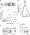
A finely tuned Ca(2+) signaling system is essential for cells to transduce extracellular stimuli, to regulate growth, and to differentiate. We have recently cloned CaT-like (CaT-L), a highly selective Ca(2+) channel closely related to the epithelial calcium channels (ECaC) and the calcium transport protein CaT1. CaT-L is expressed in selected exocrine tissues, and its expression also strikingly correlates with the malignancy of prostate cancer. The expression pattern and selective Ca(2+) permeation properties suggest an important function in Ca(2+) uptake and a role in tumor progression, but not much is known about the regulation of this subfamily of ion channels. We now demonstrate a biochemical and functional mechanism by which cells can control CaT-L activity. CaT-L is regulated by means of a unique calmodulin binding site, which, at the same time, is a target for protein kinase C-dependent phosphorylation. We show that Ca(2+)-dependent calmodulin binding to CaT-L, which facilitates channel inactivation, can be counteracted by protein kinase C-mediated phosphorylation of the calmodulin binding site.
Figures


Similar articles
-
Regulation of the mouse epithelial Ca2(+) channel TRPV6 by the Ca(2+)-sensor calmodulin.J Biol Chem. 2004 Jul 9;279(28):28855-61. doi: 10.1074/jbc.M313637200. Epub 2004 Apr 30. J Biol Chem. 2004. PMID: 15123711
-
Enkurin is a novel calmodulin and TRPC channel binding protein in sperm.Dev Biol. 2004 Oct 15;274(2):426-35. doi: 10.1016/j.ydbio.2004.07.031. Dev Biol. 2004. PMID: 15385169
-
Calmodulin and calcium interplay in the modulation of TRPC5 channel activity. Identification of a novel C-terminal domain for calcium/calmodulin-mediated facilitation.J Biol Chem. 2005 Sep 2;280(35):30788-96. doi: 10.1074/jbc.M504745200. Epub 2005 Jun 29. J Biol Chem. 2005. PMID: 15987684
-
Epithelial Ca2+ entry channels: transcellular Ca2+ transport and beyond.J Physiol. 2003 Sep 15;551(Pt 3):729-40. doi: 10.1113/jphysiol.2003.043349. Epub 2003 Jul 17. J Physiol. 2003. PMID: 12869611 Free PMC article. Review.
-
Regulation of voltage-gated Ca2+ channels by calmodulin.Sci STKE. 2005 Dec 20;2005(315):re15. doi: 10.1126/stke.3152005re15. Sci STKE. 2005. Corrected and republished in: Sci STKE. 2006 Jan 17;2006(318):er1. doi: 10.1126/stke.3182006er1. PMID: 16369047 Corrected and republished. Review.
Cited by
-
The nociceptor ion channel TRPA1 is potentiated and inactivated by permeating calcium ions.J Biol Chem. 2008 Nov 21;283(47):32691-703. doi: 10.1074/jbc.M803568200. Epub 2008 Sep 5. J Biol Chem. 2008. PMID: 18775987 Free PMC article.
-
Active intestinal calcium transport in the absence of transient receptor potential vanilloid type 6 and calbindin-D9k.Endocrinology. 2008 Jun;149(6):3196-205. doi: 10.1210/en.2007-1655. Epub 2008 Mar 6. Endocrinology. 2008. PMID: 18325990 Free PMC article.
-
Interplay between calmodulin and phosphatidylinositol 4,5-bisphosphate in Ca2+-induced inactivation of transient receptor potential vanilloid 6 channels.J Biol Chem. 2013 Feb 22;288(8):5278-90. doi: 10.1074/jbc.M112.409482. Epub 2013 Jan 8. J Biol Chem. 2013. PMID: 23300090 Free PMC article.
-
Excision of Trpv6 gene leads to severe defects in epididymal Ca2+ absorption and male fertility much like single D541A pore mutation.J Biol Chem. 2012 May 25;287(22):17930-41. doi: 10.1074/jbc.M111.328286. Epub 2012 Mar 15. J Biol Chem. 2012. PMID: 22427671 Free PMC article.
-
Structure-function analysis of TRPV channels.Naunyn Schmiedebergs Arch Pharmacol. 2005 Apr;371(4):285-94. doi: 10.1007/s00210-005-1053-7. Naunyn Schmiedebergs Arch Pharmacol. 2005. PMID: 15889240 Review.
References
-
- Clapham D E. Cell. 1995;80:259–268. - PubMed
-
- Kohn E C, Liotta L A. Cancer Res. 1995;55:1856–1862. - PubMed
-
- Hoenderop J G, van der Kemp A W, Hartog A, van de Graaf S F, Van Os CH, Willems P H, Bindels R J. J Biol Chem. 1999;274:8375–8378. - PubMed
-
- Peng J B, Chen X Z, Berger U V, Vassilev P M, Tsukaguchi H, Brown E M, Hediger M A. J Biol Chem. 1999;274:22739–22746. - PubMed
Publication types
MeSH terms
Substances
LinkOut - more resources
Full Text Sources
Other Literature Sources
Molecular Biology Databases
Miscellaneous

