

Transforming growth factor-beta and breast cancer: Mammary gland development
- PMID: 11250698
- PMCID: PMC139430
- DOI: 10.1186/bcr40
Transforming growth factor-beta and breast cancer: Mammary gland development
Abstract
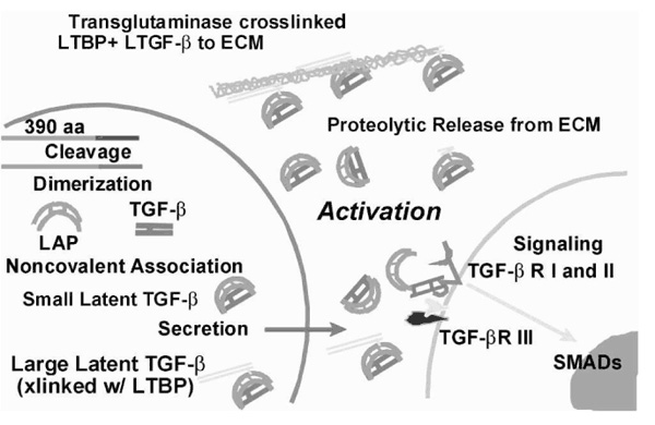
Transforming growth factor (TGF)-beta1 is a pluripotent cytokine that profoundly inhibits epithelial proliferation, induces apoptosis, and influences morphogenesis by mediating extracellular matrix deposition and remodeling. The physiologic roles of the action of TGF-beta in mammary gland, indeed in most tissues, are poorly understood. In order to understand the actions of TGF-beta, we need to take into account the complexity of its effects on different cell types and the influence of context on cellular responses. This task is further compounded by multiple mechanisms for regulating TGF-beta transcription, translation, and activity. One of the most significant factors that obscures the action of TGF-beta is that it is secreted as a stable latent complex, which consists of the 24-kDa cytokine and the 80-kDa dimer of its prepro region, called latency-associated peptide. Latency imposes a critical restraint on TGF-beta activity that is often overlooked. The extracellular process known as activation, in which TGF-beta is released from the latent complex, is emphasized in the present discussion of the role of TGF-beta in mammary gland development. Definition of the spatial and temporal patterns of latent TGF-beta activation in situ is essential for understanding the specific roles that TGF-beta plays during mammary gland development, proliferation, and morphogenesis.
Figures
References
-
- Daniel CW, Robinson SD. Regulation of mammary growth and function by TGF-β. . Mol Reprod Dev. 1992;32:145–151. - PubMed
-
- Knabbe C, Lippman ME, Wakefield LM, et al. Evidence that transforming growth factor-β is a hormonally regulated negative growth factor in human breast cancer cells. Cell. 1987;48:417–428. - PubMed
-
- Wakefield l, Colletta AA, McCune BK, Sporn MB. Roles for transforming growth factors-β in the genesis, prevention and treatment of breast cancer. Genes, Oncogens, and Hormones: Advances in Cellular and Molecular Biology of Breast Cancer. Edited by Dickson RB, Lipman MF. Boston: Kluwer Academic Publishers, 1991. pp. 97–136.
-
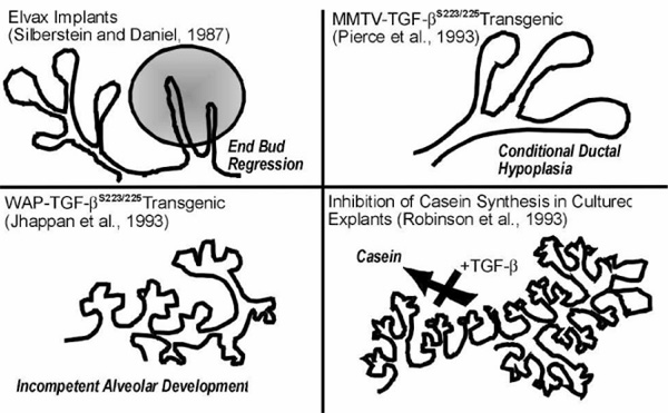
- Silberstein GB, Daniel CW. Reversible inhibition of mammary gland growth by transforming growth factor-β. . Science. 1987;237:291–293. - PubMed
-
- Daniel CW, Silberstein GB, Van Horn K, Strickland P, Robinson S. TGF-β1-induced inhibition of mouse mammary ductal growth: developmental specificity and characterization. . Dev Biol. 1989;135:20–30. - PubMed
Publication types
MeSH terms
Substances
LinkOut - more resources
Full Text Sources
Other Literature Sources
Medical
