Review
. 2000;2(5):345-52.
doi: 10.1186/bcr79. Epub 2000 Jul 12.

Estrogen receptor transcription and transactivation: Estrogen receptor knockout mice: what their phenotypes reveal about mechanisms of estrogen action

Affiliations
Review

Estrogen receptor transcription and transactivation: Estrogen receptor knockout mice: what their phenotypes reveal about mechanisms of estrogen action

S Curtis Hewitt et al. Breast Cancer Res. 2000.

Abstract

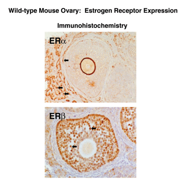
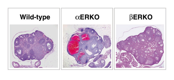
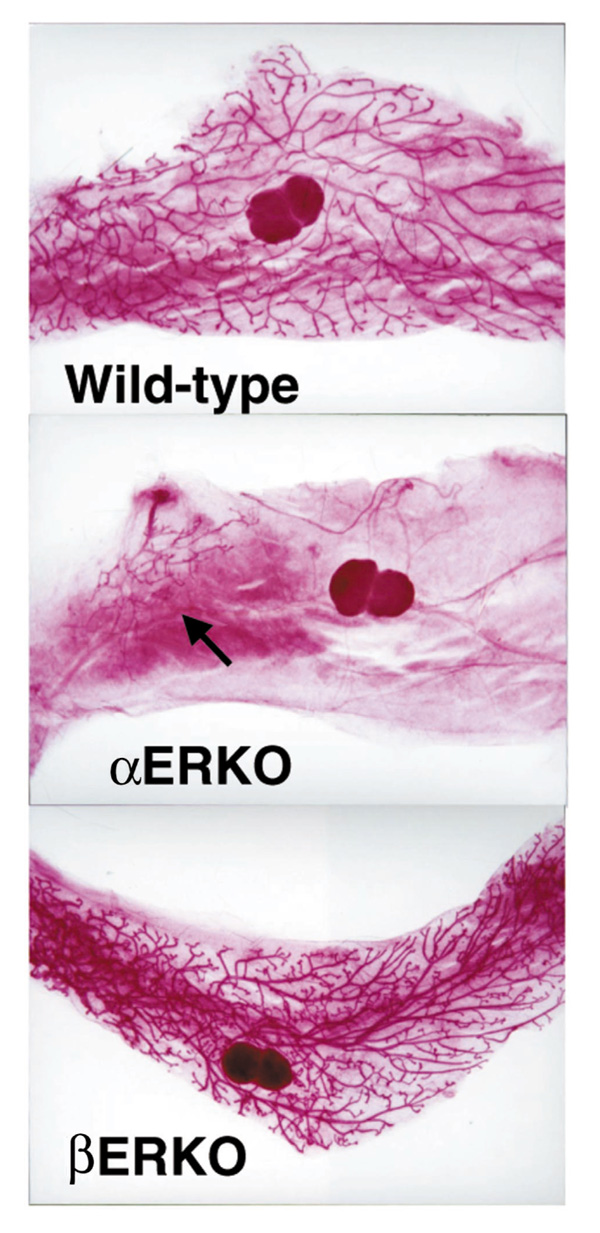
Natural, synthetic and environmental estrogens have numerous effects on the development and physiology of mammals. Estrogen is primarily known for its role in the development and functioning of the female reproductive system. However, roles for estrogen in male fertility, bone, the circulatory system and immune system have been established by clinical observations regarding sex differences in pathologies, as well as observations following menopause or castration. The primary mechanism of estrogen action is via binding and modulation of activity of the estrogen receptors (ERs), which are ligand-dependent nuclear transcription factors. ERs are found in highest levels in female tissues critical to reproduction, including the ovaries, uterus, cervix, mammary glands and pituitary gland. Since other affected tissues have extremely low levels of ER, indirect effects of estrogen, for example induction of pituitary hormones that affect the bone, have been proposed. The development of transgenic mouse models that lack either estrogen or ER have proven to be valuable tools in defining the mechanisms by which estrogen exerts its effects in various systems. The aim of this article is to review the mouse models with disrupted estrogen signaling and describe the associated phenotypes.


Figures

Figure 1
Figure 1
Pathology of adult αERKO and βERKO testes. Sections from wild-type and ER-disrupted testes were stained with hematoxylin and eosin for comparison of their pathology. The wild-type and βERKO testes are indistinguishable, while the αERKO testis shows degeneration of the testicular structures.
Figure 2
Figure 2
Gross morphology of adult ERKO female reproductive tracts. Reproductive tracts dissected from wild-type and βERKO animals are normal, while the αERKO uterus is immature and the ovaries are enlarged and dark-colored due to hemorrhagic cysts.
Figure 3
Figure 3
Uterine and vaginal histology of adult ERKO mice. Histological analysis of the uterus (top panels) and vagina (bottom panels) shows the βERKO tissue is indistinguishable from the wild type, showing the normal organization of the uterine tissue into the epithelial (Ep), stromal (St) and myometrial (My) compartments. In contrast, the αERKO uterine tissue is composed of all three layers, yet they are all immature and hypoplastic. Note also the glands are fewer in number. The vagina of the wild-type and βERKO mice is identical and cornified, indicating response to estrogen, while the αERKO vagina shows no cornification. (Reproduced with permission from Couse and Korach [5**].)
Figure 4
Figure 4
Localization of ERα and ERβ protein in the ovary. Serial sections from mouse ovary were stained with antibody to ERα (top panel) or ERβ (bottom panel). Note that ERβ immunoreativity is confined to the granulosa cells of the follicle, while ERα immunoreativity is localized in the thecal and interstitial cells of the ovary.
Figure 5
Figure 5
Ovarian pathology of the ERKO mice. Histological analysis of the wild-type ovary shows normal follicular development and indications of ovulation. The αERKO ovary shows large cystic structures and arrested follicle development with no indication of ovulation, while the βERKO ovary shows development of follicles is occurring but with little indication of successful ovulation.
Figure 6
Figure 6
Mammary gland whole mounts from ERKO mice. An adult βERKO mouse displays a fully developed ductal network similar to the wild type. In contrast, the αERKO mouse has only a rudimentary underdeveloped epithelial duct (arrow).

References

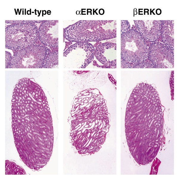
    1. Eddy EM, Washburn TF, Bunch DO, Goulding EH, Gladen BC, Lubahn DB, Korach KS. Targeted disruption of the estrogen receptor gene in male mice causes alteration of spermatogenesis and infertility. . Endocrinology. 1996;137:4796–4805. - PubMed
    1. Hess RA, Bunick D, Lee KH, Bahr J, Taylor JA, Korach KS, Lubahn DB. A role for oestrogens in the male reproductive system. . Nature. 1997;390:509–512. - PMC - PubMed
    1. Mahato D, Goulding EH, Korach KS, Eddy EM. Spermatogenic cells do not require estrogen receptor alpha for development or function. . Endocrinology. 2000;141:1273–1276. - PubMed
    1. Lubahn DB, Moyer JS, Golding TS, Couse JF, Korach KS, Smithies O. Alteration of reproductive function but not prenatal sexual development after insertional disruption of the mouse estrogen receptor gene. . Proc Natl Acad Sci USA. 1993;90:11162–11166. - PMC - PubMed
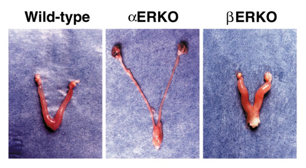
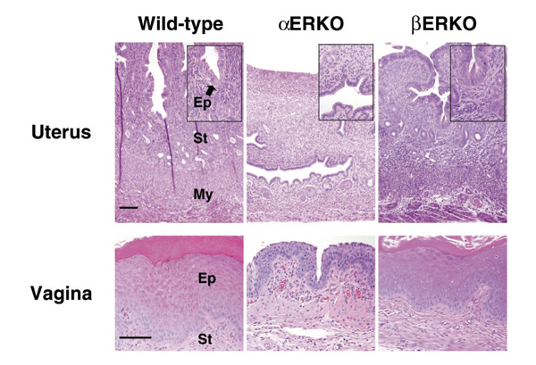
    1. Couse JF, Korach KS. Estrogen receptor null mice: what have we learned and where will they lead us? Endocr Rev. 1999;20:358–417. - PubMed

Publication types

LinkOut - more resources