Review
. 2001;3(1):28-35.
doi: 10.1186/bcr267. Epub 2000 Dec 11.

Recent advances in imaging endogenous or transferred gene expression utilizing radionuclide technologies in living subjects: applications to breast cancer

Affiliations
Review

Recent advances in imaging endogenous or transferred gene expression utilizing radionuclide technologies in living subjects: applications to breast cancer

F Berger et al. Breast Cancer Res. 2001.

Abstract

A variety of imaging technologies is being investigated as tools for studying gene expression in living subjects. Two technologies that use radiolabeled isotopes are single photon emission computed tomography (SPECT) and positron emission tomography (PET). A relatively high sensitivity, a full quantitative tomographic capability, and the ability to extend small animal imaging assays directly into human applications characterize radionuclide approaches. Various radiolabeled probes (tracers) can be synthesized to target specific molecules present in breast cancer cells. These include antibodies or ligands to target cell surface receptors, substrates for intracellular enzymes, antisense oligodeoxynucleotide probes for targeting mRNA, probes for targeting intracellular receptors, and probes for genes transferred into the cell. We briefly discuss each of these imaging approaches and focus in detail on imaging reporter genes. In a PET reporter gene system for in vivo reporter gene imaging, the protein products of the reporter genes sequester positron emitting reporter probes. PET subsequently measures the PET reporter gene dependent sequestration of the PET reporter probe in living animals. We describe and review reporter gene approaches using the herpes simplex type 1 virus thymidine kinase and the dopamine type 2 receptor genes. Application of the reporter gene approach to animal models for breast cancer is discussed. Prospects for future applications of the transgene imaging technology in human gene therapy are also discussed. Both SPECT and PET provide unique opportunities to study animal models of breast cancer with direct application to human imaging. Continued development of new technology, probes and assays should help in the better understanding of basic breast cancer biology and in the improved management of breast cancer patients.


Figures

Supplementary Figure 1
Schematic illustrating single photon emission computed tomography (SPECT) and positron emission tomography (PET). (a) In SPECT, a single photon is produced as the isotope decays and this single photon must be detected through rotating detectors using a collimator. (b) In PET, annihilation eventually occurs with the positron and electron to produce two high energy (511 keV) gamma rays at ~ 180° that are detected using a circular ring of detectors.
Supplementary Figure 2
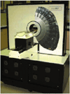
Photograph of the microPET scanner designed at UCLA, Los Angeles, CA, USA. The scanner is capable of imaging small animals such as mice in 30-60 min with a spatial resolution of ~ 2 mm in each axis (8 mm3). The scanner uses an 8 × 8 array of 2 × 2 × 10 mm3 lutetium oxyorthosilicate crystals. The ring diameter is 17.1 cm. The transaxial and axial fields of view are 17.1 and 1.8 cm, respectively.
Figure 1
Figure 1
MicroPET imaging of a mouse using a [64Cu]-labeled antibody fragment targeted against carcinoembryonic antigen (CEA) in a xenograft model. The mouse carried a C6 glioma xenograft on the left shoulder (negative control) and a LS174T xenograft expressing CEA on the right shoulder (arrows). The mouse was injected with 26 μCi 64Cu-anti-CEA minibody and imaged at 5 h, with the highest retention in the LS174T tumor (arrow) and liver, and lower retention in the control tumor (arrowhead). The liver signal occurs due to metabolism of the antibody fragment at that site.
Figure 2
Figure 2
Illustration of antisense hybridization imaging approach. Small radioactive antisense oligodeoxynucleotides (RASONs) are used to target a small portion of a chosen mRNA. If sufficient levels of mRNA are present, it may be possible to retain the RASONs only in those cells expressing the mRNA. Efflux would occur in other cells if non-specific interactions can be minimized. Because radiolabeling chemistry can be made independent of the RASON sequence, many different types of mRNA can potentially be targeted.
Figure 3
Figure 3
Schematic illustrating a reporter gene approach. An imaging reporter gene has to be introduced into the cell, which is driven by a promoter of choice. Transcription of the imaging reporter gene with subsequent translation of the mRNA leads to an enzyme. This enzyme can selectively trap an imaging reporter probe. The imaging reporter probe will not be trapped in those cells in which there is no expression of the imaging reporter gene. Note that it is also possible for the imaging reporter gene to encode for an intracellular and/or cell surface receptor. This receptor would then bind the imaging reporter probe (a ligand). Levels of the trapped probe can be related to levels of imaging reporter gene expression in either approach.
Figure 4
Figure 4
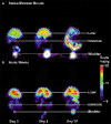
Adenoviral-mediated HSV1-sr39tk gene expression repetitively imaged in two mice. Each mouse was tail-vein injected with 1.0 × 10⁹ pfu adenovirus in which the HSV1-sr39tk reporter gene is driven by the CMV promoter. Each mouse was imaged in a microPET system at the days indicated 1 h after tail-vein injection of 250 μCi [18F]-labeled penciclovir molecule (FHBG). The scale represents the accumulation of FHBG measured as percent injected dose per gram of tissue (%ID/g). (a) The Swiss Webster mouse shows a decrease in gene expression over time, and (b) the nude mouse shows persistence of gene expression in the liver. These differences are likely due to the immune system being fully competent in the Swiss Webster mouse.
Supplementary Figure 3
Imaging of two reporter genes in the same mouse. A nude mouse carrying two tumors (left, dopamine 2 receptor [D2R]; right, herpes simplex virus type 1 thymidine kinase [HSV1-tk]) was imaged in a microPET system with tail-vein injection of ~ 250 μCi [18F]-fluoroethylspiperone (FESP) and, 24 hours later, with ~ 250 μCi 8-[18F]-fluoropenciclovir (FPCV). Imaging began 1 hour after injection of each tracer. These results show the ability to image both the D2R and the HSV1-tk reporter genes in the same living mouse with microPET. The scale represents the accumulation of FESP and FPCV measured as percent injected dose per gram of tissue (%ID/g).
Supplementary Figure 4
Imaging bicistronic gene expression. MicroPET imaging of the pCMV-D2R-IRES-tkm C6 tumors in a nude mouse. Three C6 cell lines stably transfected with pCMV-D2R-IRES-sr39tk (labeled A, B, C) and the C6 parental cell line (labeled D) were injected into four separate sites in a single mouse. After 10 days, when each tumor was at least 6 mm in diameter, the mouse was imaged with 2-[18F]-2-fluoro-deoxyglucose (FDG), followed 24 h later with [18F]-fluoroethylspiperone (FESP), followed 24 h later with 8-[18F]-fluoropenciclovir (FPCV). The FDG (whole body) image represents the average of all coronal (horizontal) planes and therefore the four tumors are not well visualized. The FDG (section) image taken from a set of planar images passing through all four tumors shows accumulation of FDG in all four tumors. Correlated signal intensity is observed between the FESP and FPCV images for each tumor. There is a high correlation between FESP and FPCV microPET signals. Br, Brain; Bl, bladder; R, rectum; %ID/g, percent injected dose per gram of tissue.

References

    1. Budinger TF. Critical review of PET, SPECT and neuroreceptor studies in schizophrenia. J Neural Transm. 1992;36(suppl):3–12. - PubMed
    1. Phelps ME. Inaugural article: positron emission tomography provides molecular imaging of biological processes. Proc Natl Acad Sci USA. 2000;97:9226–9233. - PMC - PubMed
    1. Fleming JS, Alaamer AS. Influence of collimator characteristics on quantification in SPECT. J Nucl Med. 1996;37:1832–1836. - PubMed
    1. Freifelder R, Karp JS. Dedicated PET scanners for breast imaging. Phys Med Biol. 1997;42:2463–2480. doi: 10.1088/0031-9155/42/12/012. - DOI - PubMed
    1. Cherry SR, Shao Y, Silverman RW, Meadors K, Siegel S, Chatziioannou A, Young JW, Jones WF, Moyers JC, Newport D, Boutefnouchet A, Farquhar TH, Andreaco M, Paulus MJ, Binkley DM, Nutt R, Phelps ME. MicroPET: a high resolution PET scanner for imaging small animals. IEEE Trans Nucl Sci. 1997;44:1161–1166. doi: 10.1109/23.596981. - DOI

Publication types