. 2001 Mar 15;20(6):1215-22.
doi: 10.1093/emboj/20.6.1215.

Multiple classes of stem cells in cutaneous epithelium: a lineage analysis of adult mouse skin

Affiliations

Multiple classes of stem cells in cutaneous epithelium: a lineage analysis of adult mouse skin

S Ghazizadeh et al. EMBO J. .

Abstract

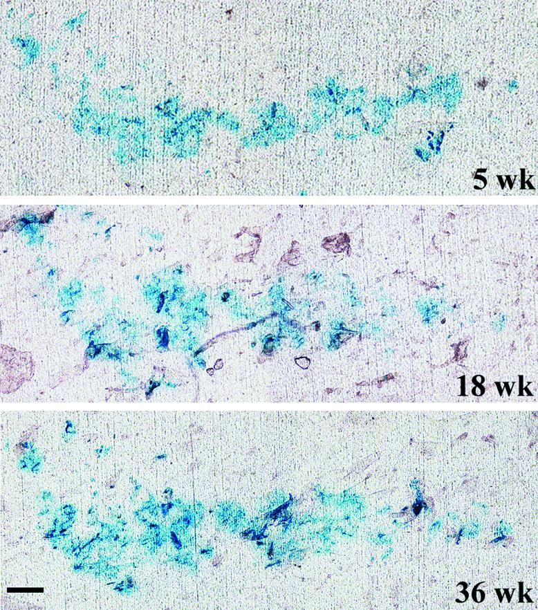
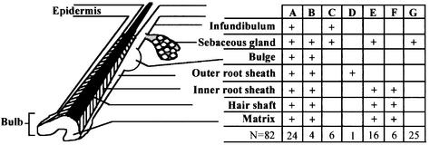
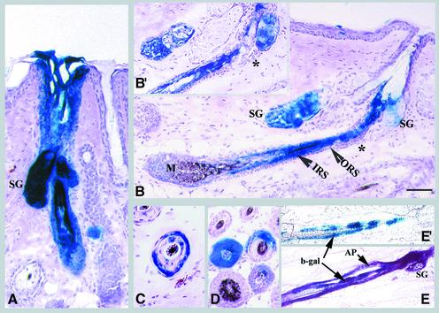
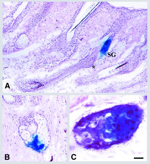
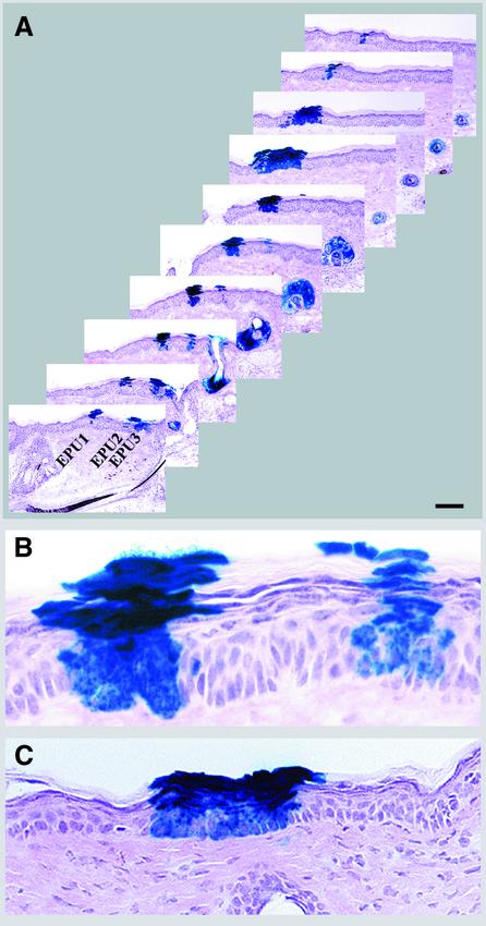
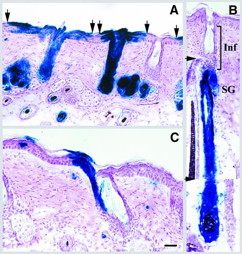
Continuous renewal of the epidermis and its appendages throughout life depends on the proliferation of a distinct population of cells called stem cells. We have used in situ retrovirus-mediated gene transfer to genetically mark cutaneous epithelial stem cells of adolescent mice, and have followed the fate of the marked progeny after at least 37 epidermal turnovers and five cycles of depilation-induced hair growth. Histological examination of serial sections of labeled pilosebaceous units demonstrated a complex cell lineage. In most instances, labeled cells were confined to one or more follicular compartments or solely to sebaceous glands. Labeled keratinocytes in interfollicular epidermis were confined to distinct columnar units representing epidermal proliferative units. The contribution of hair follicles to the epidermis was limited to a small rim of epidermis at the margin of the follicle, indicating that long term maintenance of interfollicular epidermis was independent of follicle-derived cells. Our results indicate the presence of multiple stem cells in cutaneous epithelium, some with restricted lineages in the absence of major injury.


Figures

None
Fig. 1. Persistent expression of β-gal in epidermis. Dorsal skin of a MtnlacZ transgenic mouse was transduced with retroviruses encoding β-gal and, at the times indicated, β-gal expression was assessed by tape stripping and staining of adherent cornified cells with X-gal. Bar = 100 µm.
None
Fig. 2. Distribution of lacZ-positive cells in various compartments of labeled pilosebaceous units. Serial sections of skin of transduced mice following five cycles of induced hair growth, at 37 weeks post-transduction, were analyzed by X-gal staining and the distribution of lacZ-positive clusters in the various compartments of 82 pilosebaceous units was noted. ‘+’ indicates partial or uniform labeling of cells in that compartment. The number of labeled pilosebaceous units in each category is shown in the bottom row.
None
Fig. 3. Lineage analysis of hair follicles. (A–D) Mice were treated as described in Figure 2. Representative longitudinal sections are shown of (A) an anagen follicle with uniform labeling of all compartments, including sebaceous gland (SG) (the section shown does not show the full length of the follicle), (B) an anagen follicle with distinct labeling in the IRS and hair shaft in the absence of staining in the ORS, including the bulge (*) and (B′) an adjacent section of (B) demonstrating the absence of β-gal-positive cells in the bulge (*). Note that labeled cells in (B) are traced back to hair matrix (M). The lower panels show follicles with β-gal-positive cells confined mainly to the ORS (C) or to a portion of follicular compartments (D). (E and E′) Rag-2 mice were transduced with a mixture of two viruses encoding PLAP and β-gal. Sections were stained for PLAP (AP, purple) and β-gal (blue). (E′) is an adjacent section of (E) that was stained with X-gal alone to demonstrate confinement of β-gal-positive cells to one compartment. Scale bar: 60 µm (A and C); 100 µm (B and D); 120 µm (E and E′).
None
Fig. 4. Presence of long-lived progenitor cells in sebaceous glands. The experiment was the same as Figure 2. In (A), representative longitudinal sections of follicles with β-gal-positive cells confined solely to sebaceous glands are shown. Note the absence of β-gal-positive cells in the bulge (*). Representative cross-sections of a partially stained sebaceous gland (B) or a transduced gland with distinct clusters of β-gal- (blue) and alkaline phosphatase-positive (purple) cells (C) are shown. Scale bars: 50 µm (A); 35 µm (B); 5 µm (C).
None
Fig. 5. Lineage analysis of mouse epidermis. (A–C) The experiment was the same as Figure 2. (A) Every third section of serially cut transduced epidermis (extending over a 210 µm thickness) is presented to reveal the origin of EPUs (follicular or interfollicular). Note the different sizes of EPU1 and 2. EPU3 is an extension of follicular infundibulum. (B) Higher magnification of the fifth section of interfollicular epidermis shown in (A). (C) Follicular extensions of the follicle shown in Figure 3A appear as an EPU in adjacent sections. The distance between sections shown here and that shown in Figure 3A is 42 µm. Scale bar: 150 µm (A); 25 µm (B); 35 µm (C).
None
Fig. 6. Re-epithelialization of abraded epidermis by epithelial cells in infundibulum. Transduced skin was abraded for a second time at 36 weeks post-transduction, and 6 weeks later serial sections of skin were analyzed. (A) The increased size of follicular extensions indicates re-epithelialization by follicle-derived cells. Arrows denote the boundary of follicular extensions. (B) A composite of three serial sections to illustrate labeling of only the lower portion of a follicle. The arrowheads denote the photo splice sites. The non-labeled infundibulum (Inf) is shown. (C) The upper portion of a follicle labeled in one side of infundibulum demonstrates extension of labeled cells to epidermis. The isolated X-gal-positive cells in the dermis are transduced fibroblasts. Scale bar: 60 µm (A–B); 40 µm (C).

References

    1. Al-Barwari S.E. and Potten,C.S. (1976) Regeneration and dose–response characteristics of irradiated mouse dorsal epidermal cells. Int. J. Radiat. Biol. Relat. Stud. Phys. Chem. Med., 30, 201–216. - PubMed
    1. Allen T.D. and Potten,C.S. (1974) Fine-structural identification and organization of the epidermal proliferative unit. J. Cell Sci., 15, 291–319. - PubMed
    1. Argyris T.S. (1976) Kinetics of epidermal production during epidermal regeneration following abrasion in mice. Am. J. Pathol., 83, 329–340. - PMC - PubMed
    1. Burns J.C., Friedmann,T., Driever,W., Burrascano,M. and Yee,J.K. (1993) Vesicular somatitis virus G glycoprotein pseudotyped retroviral vectors: concentration to very high titer and efficient gene transfer into mammalian and non-mammalian cells. Proc. Natl Acad. Sci. USA, 90, 8033–8037. - PMC - PubMed
    1. Cepko C.L., Ryder,E., Austin,C., Golden,J., Fields-Berry,S. and Lin,J. (1998). Lineage analysis using retroviral vectors. Methods, 14, 393–406. - PubMed

Publication types