. 2001 Mar 15;20(6):1245-58.
doi: 10.1093/emboj/20.6.1245.

Enteropathogenic Escherichia coli mediates antiphagocytosis through the inhibition of PI 3-kinase-dependent pathways

Affiliations

Enteropathogenic Escherichia coli mediates antiphagocytosis through the inhibition of PI 3-kinase-dependent pathways

J Celli et al. EMBO J. .

Abstract

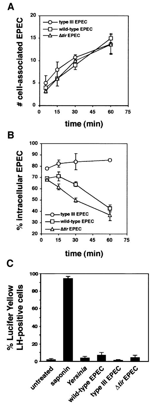
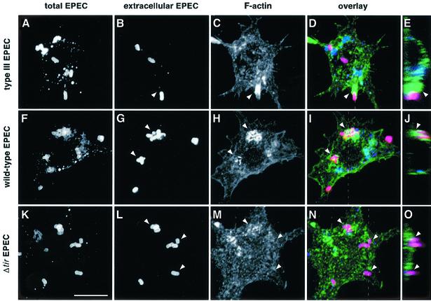
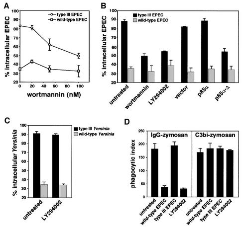
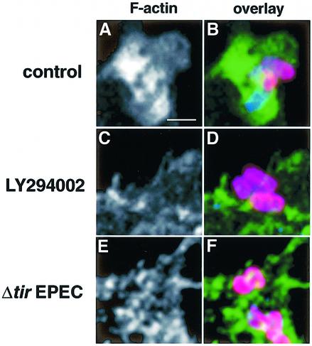
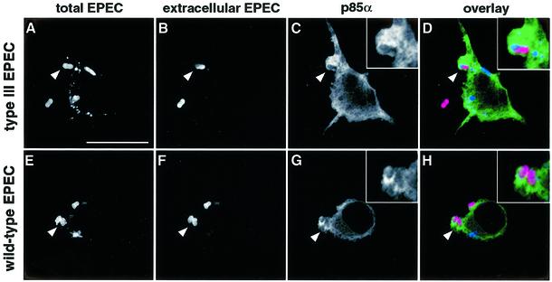
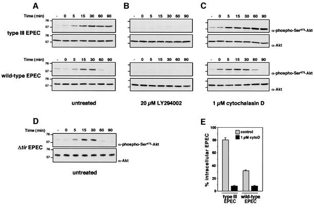
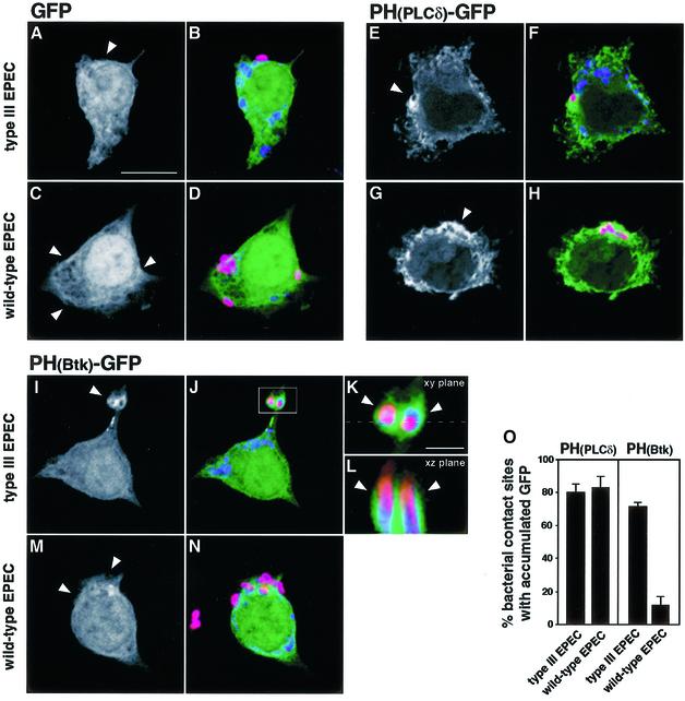
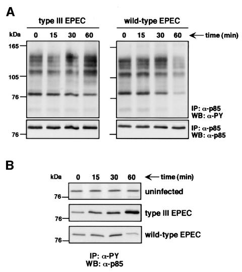
The extracellular pathogen enteropathogenic Escherichia coli (EPEC) uses a type III secretion system to inhibit its uptake by macrophages. We show that EPEC antiphagocytosis is independent of the translocated intimin receptor Tir and occurs by preventing F-actin polymerization required for bacterial uptake. EPEC-macrophage contact triggered activation of phosphatidylinositol (PI) 3-kinase, which was subsequently inhibited in a type III secretion-dependent manner. Inhibition of PI 3-kinase significantly reduced uptake of a secretion-deficient mutant, without affecting antiphagocytosis by the wild type, suggesting that EPEC blocks a PI 3-kinase-dependent phagocytic pathway. EPEC specifically inhibited Fc gamma receptor- but not CR3-receptor mediated phagocytosis of opsonized zymosan. We showed that EPEC inhibits PI 3-kinase activity rather than its recruitment to the site of bacterial contact. Phagocytosis of a secretion mutant correlated with the association of PI 3-kinase with tyrosine-phosphorylated proteins, which wild-type EPEC prevented. These results show that EPEC blocks its uptake by inhibiting a PI 3-kinase-mediated pathway, and translocates effectors other than Tir to interfere with actin-driven host cell processes. This constitutes a novel mechanism of phagocytosis avoidance by an extracellular pathogen.


Figures

None
Fig. 1. Activated EPEC establishes Tir-independent antiphagocytosis within 60 min after interaction with LMmev macrophages. Macrophages were infected with early logarithmic phase wild-type, type III secretion-defective or Δtir EPEC strains for the indicated times. Samples were processed for immunofluorescent differential EPEC staining, and the number of cell-associated EPEC (A) and the percentage of intracellular bacteria (B) were determined as described in Materials and methods. (C) Lucifer Yellow exclusion assay on cells infected with Y.enterocolitica E 40 wild-type strain, the wild-type, type III secretion-deficient or Δtir EPEC strains shows no cytoxicity associated with Yersinia or EPEC antiphagocytosis. Treatment of cells with 0.005% saponin was used as positive control of pore formation. Data are means ± SEM from three to five independent experiments.
None
Fig. 2. F-actin rearrangements at the site of EPEC adhesion to macrophages. LMmev macrophages were infected for 60 min with the secretion-deficient (A–E), wild-type (F–J) or the Δtir EPEC (K–O) strains. Samples were processed for confocal microscopy. (A, F and K) Staining of total cell-associated EPEC using a rabbit polyclonal anti-EPEC antiserum and Cy5-conjugated donkey anti-rabbit antibodies. (B, G and L) Staining of extracellular EPEC adhering to macrophages using a rabbit polyclonal anti-EPEC antiserum and Alexa™594-conjugated goat anti-rabbit antibodies. (C, H and M) F-actin staining using Alexa™488-conjugated phalloidin. (D, I and N). Three colour images obtained by merging the total bacteria, extracellular bacteria and F-actin projections. (E, J and O) Z cuts along the planes indicated by the dashed lines of (D), (I) and (N), respectively. Extracellular bacteria appear in pink, intracellular bacteria in blue, and F-actin in green. F-actin rearrangements underneath extracellular adhering bacteria are shown by arrowheads. Scale bar, 10 µm.
None
Fig. 3. Phagocytosis of secretion-deficient EPEC requires PI 3-kinase. (A) Effect of wortmannin on EPEC phagocytosis. Macrophages were treated with increasing concentrations of wortmannin, then infected for 60 min with either the secretion-deficient (type III EPEC) or the secretion-competent (wild-type EPEC) strains. Data are means ± SEM of the percentage of intracellular bacteria from three independent experiments. (B) LMmev macrophages were pre-treated with PI 3-kinase inhibitors wortmannin (100 nM) or LY294002 (25 µM), or transiently transfected with a tagged pEF-BOS vector expressing the rat CD2, with pEF-BOS-derived constructs expressing native or dominant-negative forms of p85α for 18 h. Macrophages were then infected for 60 min. Transfected cells were detected using mouse monoclonal anti-CD2 antibodies for vector transfectants and mouse monoclonal anti-p85α antibodies for p85α or p85α-Δ transfectants. The percentage of intracellular bacteria was scored only for transfected macrophages. (C) Phagocytosis assays of wild-type and type III secretion-deficient Y.enterocolitica strains in untreated or 25 µM LY294002-treated macrophages. Cells were infected for 40 min and phagocytosis was analysed as described in Materials and methods. (D) Effect of EPEC infection on phagocytosis of IgG- or C3bi-opsonized zymosan by macrophages. Cells were infected with EPEC for 60 min, or treated with 25 µM LY294002, then challenged with either IgG- or C3bi-opsonized zymosan as described in Materials and methods. All data are means ± SEM from 3–5 independent experiments.
None
Fig. 4. Inhibition of PI 3-kinase activity prevents F-actin polymerization at the site of secretion-deficient EPEC contact. LMmev macrophages were treated with 25 µM LY294002 and infected for 60 min with the type III secretion EPEC mutant. Control represents cells with no inhibitor treatment. Samples were processed for confocal microscopy as described in Materials and methods. (A and B) Details of an untreated macrophage phagocytosing bacteria. Intracellular (blue) and extracellular bacteria (pink) are surrounded by massive F-actin polymerization (green). (C and D) Details of a LY294002-treated macrophage with extracellular adhering bacteria (pink). (E and F) Details of a Δtir EPEC-infected macrophage showing no F-actin polymerization underneath extracellular bacteria (pink). Scale bar, 1 µm.
None
Fig. 5. PI 3-kinase is recruited to the site of EPEC contact. LMmev macrophages were transiently transfected with a construct overexpressing the regulatory subunit of PI 3-kinase p85α. After 18 h transfection, cells were infected for 60 min with either the secretion-deficient (type III EPEC, A–D) or the secretion-competent (wild-type EPEC, E–H) EPEC strains. Samples were processed for confocal microscopy to localize PI 3-kinase. p85α-overexpressing cells were detected using mouse monoclonal anti-p85α followed by Alexa™488-conjugated goat anti-mouse antibodies. Total and extracellular EPEC were labelled as described in Figure 2. (AD) A secretion-deficient bacterium (arrowhead) in the process of being phagocytosed (intracellular half blue, extracellular half pink; see close-up in D) is surrounded by dense p85α staining (green; C and D). (EH) p85α staining (green; G and H) also localizes with extracellular (pink; H) secretion-competent bacteria (arrowheads, F and G). In a representative experiment, PI 3-kinase recruitment was detected for 88 and 84% of the secretion-deficient and wild-type bacteria analysed, respectively. Scale bar, 10 µm.
None
Fig. 6. EPEC inhibits contact-triggered PI 3-kinase-dependent Akt phosphorylation. LMmev macrophages were infected with different EPEC strains in time course experiments. Total cell lysates were resolved on an 8% SDS–polyacrylamide gel, transferred to nitrocellulose and probed with rabbit polyclonal anti-phospho-Ser473-Akt antibodies. Blots were stripped and reprobed with rabbit polyclonal anti-Akt antibodies to ensure equal protein loadings. Experiments were performed with untreated (A and D), 20 µM LY294002- (B) or 1 µM cytochalasin D-treated macrophages (C). Molecular masses are indicated in kDa. (E) Phagocytic assays of secretion-deficient or -competent EPEC strains in control or 1 µM cytochalasin D (cytoD)-treated macrophages after 60 min infection.
None
Fig. 7. Accumulation of PIP2 and PIP3 at the site of EPEC contact with macrophages. LMmev macrophages were transiently transfected with pEGFP-N1 (A–D), pEGFP-N1-PH(PLCδ) (E–H) and pEGFP-N1-PH(Btk) (I–N) plasmids. After 18 h transfection, cells were infected for 60 min with either the secretion-deficient (type III EPEC, A, B, E, F and I–L) or the secretion-competent (wild-type EPEC, C, D, G, H, M and N) EPEC strains. Total and extracellular EPEC were labelled as described in Figure 2, and samples were analysed subsequently by confocal microscopy. Black and white projections show GFP fluorescence alone, while three-colour images are merged projections of GFP (green), total (blue) and extracellular (red) bacteria stainings. (A and B) Projection showing no GFP signal (arrowhead, A) around an extracellular secretion-deficient mutant (pink, B). (C and D) Projection showing no GFP signal (arrowheads, C) underneath extracellular wild-type bacteria (pink, D). (E and F) Projection showing PIP2 accumulation (arrowhead, E) around a secretion-deficient EPEC being phagocytosed (pink, F). (G and H) Projection showing PIP2 accumulation (arrowhead, G) in the vicinity of extracellular wild-type bacteria (pink, H). (I and J) Projection showing PIP3 accumulation (arrowhead, I) at the site of secretion-deficient EPEC contact (pink, J). (K) Magnification from (J) detailing PIP3 accumulation (arrowheads) in phagocytic cups around secretion-deficient EPEC being phagocytosed. (L) Z cut from (K), along the plane indicated by the dashed line, showing PIP3 accumulation (arrowheads) in membrane extensions around two secretion-deficient EPEC being phagocytosed (intracellular portions in blue, extracellular portions in pink). (M and N) Projection showing the absence of PIP3 accumulation (arrowhead, M) at the site of adherence of extracellular secretion-competent bacteria (pink, N). (O) Quantitation of PIP2 [PH(PLCδ) panel] and PIP3[PH(Btk) panel] accumulation at sites of type III secretion-deficient or wild-type EPEC contact. Scale bar, 10 µm, except in K and L, 1 µm.
None
Fig. 8. PI 3-kinase association with phosphotyrosine (PY) proteins in EPEC-infected macrophages. (A) PY protein content in PI 3-kinase complexes from macrophages infected either with the secretion-deficient (type III EPEC) or the secretion-competent (wild-type EPEC) strains after the indicated times. PI 3-kinase complexes were immunoprecipitated from EPEC-infected macrophages using rabbit polyclonal anti-p85α antibodies as described in Materials and methods. Immunocomplexes were resolved on an 8% SDS–polyacrylamide gel, transferred to nitrocellulose and probed with mouse monoclonal anti-PY antibodies (upper panels). Blots were stripped and reprobed with rabbit polyclonal anti-p85α antibodies to ensure equal amounts of immunoprecipitated PI 3-kinase in the different samples (lower panels). (B) PY protein-associated PI 3-kinase in uninfected, secretion-deficient (type III EPEC) or secretion-competent (wild-type EPEC) EPEC-infected macrophages. PY proteins were immunoprecipitated using mouse monoclonal anti-PY (clone 4G10) antibody as described in Materials and methods. Immunoblots were probed with rabbit polyclonal anti-p85α antibodies. Time (min) refers to the duration of infection before immunoprecipitation.

References

    1. Araki N., Johnson,M.T. and Swanson,J.A. (1996) A role for phosphoinositide 3-kinase in the completion of macropinocytosis and phagocytosis by macrophages. J. Cell Biol., 135, 1249–1260. - PMC - PubMed
    1. Black D.S. and Bliska,J.B. (1997) Identification of p130Cas as a substrate of Yersinia YopH (Yop51), a bacterial protein tyrosine phosphatase that translocates into mammalian cells and targets focal adhesions. EMBO J., 16, 2730–2744. - PMC - PubMed
    1. Caron E. and Hall,A. (1998) Identification of two distinct mechanisms of phagocytosis controlled by different Rho GTPases. Science, 282, 1717–1721. - PubMed
    1. Celli J., Deng,W. and Finlay,B.B. (2000) Enteropathogenic Escherichia coli (EPEC) attachment to epithelial cells: exploiting the host cell cytoskeleton from the outside. Cell. Microbiol., 2, 1–9. - PubMed
    1. Cox D., Tseng,C.C., Bjekic,G. and Greenberg,S. (1999) A requirement for phosphatidylinositol 3-kinase in pseudopod extension. J. Biol. Chem., 274, 1240–1247. - PubMed

Publication types

MeSH terms