. 2001 Mar 15;20(6):1373-82.
doi: 10.1093/emboj/20.6.1373.

hRRN3 is essential in the SL1-mediated recruitment of RNA Polymerase I to rRNA gene promoters

Affiliations

hRRN3 is essential in the SL1-mediated recruitment of RNA Polymerase I to rRNA gene promoters

G Miller et al. EMBO J. .

Abstract

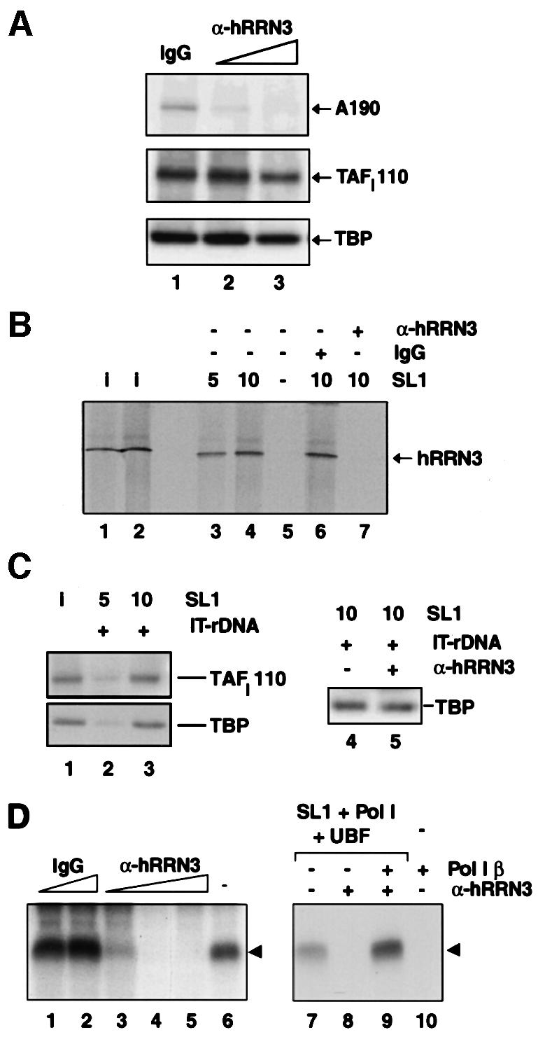
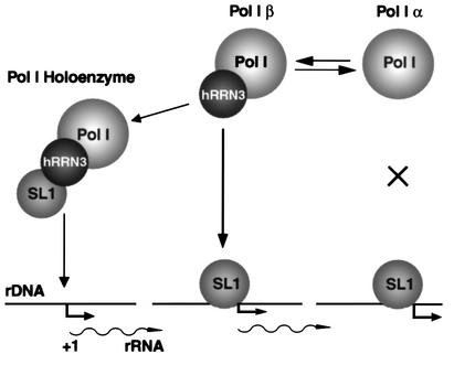
A crucial step in transcription is the recruitment of RNA polymerase to promoters. In the transcription of human rRNA genes by RNA Polymerase I (Pol I), transcription factor SL1 has a role as the essential core promoter binding factor. Little is known about the mechanism by which Pol I is recruited. We provide evidence for an essential role for hRRN3, the human homologue of a yeast Pol I transcription factor, in this process. We find that whereas the bulk of human Pol I complexes (I alpha) are transcriptionally inactive, hRRN3 defines a distinct subpopulation of Pol I complexes (I beta) that supports specific initiation of transcription. Human RRN3 interacts directly with TAF(I)110 and TAF(I)63 of promoter-selectivity factor SL1. Blocking this connection prevents recruitment of Pol I beta to the rDNA promoter. Furthermore, hRRN3 can be found in transcriptionally autonomous Pol I holoenzyme complexes. We conclude that hRRN3 functions to recruit initiation-competent Pol I to rRNA gene promoters. The essential role for hRRN3 in linking Pol I to SL1 suggests a mechanism for growth control of Pol I transcription.


Figures

None
Fig. 1. Two functionally distinct forms of human Pol I. (A) Schematic outline of the procedure for the purification of Pol I from HeLa cell nuclear extract. (B) Pol I size fractionated as a complex of >1 MDa. Samples from fractions (0.5 ml) of the Superose 6 column were tested in a specific transcription assay with the rDNA promoter, supplemented with SL1 and UBF, and transcripts were detected by S1 nuclease protection (arrowhead). The largest subunit of human Pol I, hA190, was detected in the same elution volumes from the Superose 6 column in immunoblots with anti-A190 antibodies. Size standards for the Superose 6 column are indicated above the lanes. (C) Poros Heparin columns separate Pol I α and β. The bulk of Pol I, Pol I α, is in fractions 28–32 as revealed with anti-A190 antibodies on immunoblots (bottom panel). The second peak of Pol I, Pol I β in fractions 34–36, constitutes a minor fraction of the total ‘soluble’ Pol I, yet in a reconstituted transcription assay with rDNA, SL1 and UBF support specific initiation of transcription (top panel; arrowhead). Pol I α is inactive in that same assay. (D) Mixing of Pol I α and β peak fractions. Pooled peak fractions of Pol I α and β (in µl) from the Poros Heparin column were tested, separately (lanes 1, 2 and 5) or mixed (lanes 3 and 4) as indicated above the lanes, for their ability to support specific initiation of transcription with SL1, UBF and rDNA (arrowhead).
None
Fig. 2. hRRN3 is specifically associated with promoter- and initiation-competent Pol I β. (A) Schematic outline of the purification of Pol I α and β. (B) Mono S (MS) fractions were assayed in a non-specific transcription assay with calf thymus DNA, and the activities were expressed as a percentage of maximal activity for each form of Pol I. Note that the ratio of Pol I α and β remained the same throughout the purification procedure, with Pol I α ∼10 times more abundant than Pol I β. (C) Fractions from the Mono S column for Pol I β were assayed in a reconstituted transcription assay with SL1, UBF and rDNA promoter (arrowhead). (D) Affinity-purified anti-peptide antibodies raised against hRRN3 react in immunoblots with both an Escherichia coli-expressed recombinant GST–hRRN3 fusion protein of 100 kDa (lane 1) and a single protein of 74 kDa, the predicted molecular weight for hRRN3, in HeLa cell nuclear extract (lane 5). (E) hRRN3 is specific for Pol I β, and is lacking in Pol I α. Immunoblots of peak fractions for Pol I α and Pol I β from the Mono S columns were probed with anti-human A190 (Pol I), anti-human RRN3, anti-mouse PAF53 and anti-human AC19 (subunit shared by Pol I and III) antibodies.
None
Fig. 3. hRRN3 co-localizes and co-immunoprecipitates with Pol I and is found in a complex with SL1 and UBF. (A) Imaging of EYFP–hRRN3 expression (green) in transfected HeLa cells and of the Pol I second largest subunit (A127, red), reveals co-localization in sub-nucleolar structures (merged image in the middle, where yellow indicates co-localization). Scale bar, 10 µm. (B) Extracts (2.7 mg) from HEK293 cells transfected with EYFP (lanes 1–3) and EYFP–hRRN3 (lanes 5–7) expression constructs were immunoprecipitated with anti-GFP antibodies. Immunocomplexes were subjected to SDS–PAGE, immunoblotted and probed with an antibody specific for human A190 (Pol I largest subunit). Forty micrograms of the input (lanes 1 and 5) and supernatant (sup) after the immunoprecipitations (lanes 3 and 7) were loaded, and these, therefore, represent ∼1.5% of the total protein subjected to immunoprecipitation. As a marker, we loaded highly purified Pol I (lane 4). Essentially the same immunoprecipitation results were obtained (data not shown) when the precipitations were performed in the presence of high concentrations (200 µg/ml) of ethidium bromide (Lai and Herr, 1992). (C) The immunoprecipitates of (B) were analysed for SL1 subunits with antibodies specific for TAFI63 (Comai et al., 1994; Zomerdijk et al., 1994) and a mouse monoclonal antibody, SL39, against human TBP, both at 1:1000. Input and supernatant are as in (B). Highly purified SL1 was loaded as a marker (lane 4). Note the slightly different mobilities of SL1 subunits in the relatively pure protein samples (lanes 4 and 6) compared with those in complex protein mixtures (lanes 5 and 7). (D) The immunoprecipitates of (B) were analysed in parallel for Pol I largest subunit A190, and UBF1 and 2. Anti-UBF antibodies (1:1000) were in a rabbit polyclonal serum. (E) Nuclear extracts prepared from HEK293 cells transfected with EYFP and EYFP–hRRN3 expression plasmids were tested for specific transcription initiation activity on the rDNA promoter (lanes 1 and 2). Anti-GFP-immunoprecipitated complexes from these nuclear extracts were transcriptionally active upon the addition of rRNA gene promoter template DNA and ribonucleoside triphosphates (lane 4). In the absence of added rDNA template, no transcription was observed (data not shown) and the immunoprecipitate from the EYFP-transfected cells was inactive (lane 3).
None
Fig. 4. hRRN3 interacts with SL1. (A) Highly purified SL1 (see Materials and methods) specifically interacts with recombinant and purified GST–hRRN3, as revealed by immunoblotting of the relevant strips of the immunoblot with antibodies specific for three subunits of SL1, TAFI110, TAFI63 and TAFI48. (B) hRRN3 interacts with SL1, which had been immunoprecipitated with antibodies specific for TBP. FLAG-epitope affinity-purified, 35S-radiolabelled hRRN3 (10% of input in lane 1) and luciferase (10% of input, lane 2) were incubated with SL1 immobilized via a TBP antibody to protein G–Sepharose beads (lanes 4 and 5). As an additional control, hRRN3 was added to antibody-loaded beads without SL1 (lane 3). Bound proteins were subjected to SDS–PAGE. The gel was fixed, dried and subjected to autoradiography. (C) FLAG-tag affinity-purified [35S]hRRN3 specifically interacts with two subunits of SL1, TAFI110 and TAFI63 in a far-western blot of highly purified SL1 (lane 1). The blot was probed with antibodies specific for TAFI110 (lane 2) and TAFI63 (lane 3), confirming their identity. (D) GST–hRRN3 interacts with two subunits of SL1. GST (lane 2 and 5) and GST–hRRN3 (lanes 3 and 6) on glutathione beads were incubated with in vitro translated [35S]methionine-labelled TAFI110 and TAFI63, and after extensive washing the resulting protein complexes were resolved by SDS–PAGE and autoradiography. Ten per cent of the TAFI110 and TAFI63 inputs are shown in lanes 1 and 4, respectively.
None
Fig. 5. hRRN3 is essential for the SL1-mediated recruitment of Pol I to the rDNA promoter. (A) Affinity-purified antibodies against hRRN3, in a dose-dependent manner, prevent the recruitment of Pol I β to SL1, which was pre-bound to the rDNA promoter. Immobilized rDNA promoter template DNA (IT-DNA) was pre-incubated for 30 min with highly purified SL1 and, in parallel, a 0.2 M KCl fraction from DEAE columns, named D0.2, and containing UBF and initiation-competent Pol I (Comai et al., 1992), was pre-incubated for 30 min with affinity-purified anti-hRRN3 antibodies (4 and 8 µg, lanes 2 and 3, respectively) or control sheep IgG antibodies (8 µg, lane 1). The immobilized templates were washed in TM10/0.05 M KCl to remove unbound SL1, and then added to the UBF/Pol I/antibody mixture. This reaction was incubated for a further 20 min, after which the immobilized templates were washed in TM10/0.05 M KCl. Template-bound proteins were eluted in 10 M urea at room temperature and analysed by immunoblotting with antibodies specific for human A190, TAFI110 (Comai et al., 1994; Zomerdijk et al., 1994) and TBP. (B) Antibodies specific for hRRN3 block the binding of highly purified hRRN3 to SL1 pre-bound to the rDNA promoter. Immobilized rDNA promoter template DNA was pre-incubated for 20 min with either 5 or 10 µl of highly purified SL1 (lanes 3 and 4, respectively) or without SL1 (lane 5). Templates were washed and mixed with FLAG-epitope affinity-purified, 35S-radiolabelled hRRN3. In a separate experiment, the purified 35S-radiolabelled hRRN3 had been pre-incubated for 20 min with peptide affinity-purified anti-hRRN3 antibodies (8 µg, lane 7) or IgG (8 µg, lane 6) before addition to promoter-bound SL1. The reactions were incubated for 20 min and then the templates were washed. The template-bound proteins were eluted in urea and subjected to SDS–PAGE. The gel was fixed, dried and subjected to autoradiography to reveal [35S]FLAG-hRRN3. Lanes 1 and 2 are 10 and 20%, respectively, of the input of [35S]FLAG-hRRN3. In the absence of SL1, hRRN3 did not bind the promoter DNA template (lane 5). (C) The binding of SL1 to the immobilized template (IT-rDNA) from the experiment described in (B) was verified by immunoblotting the reactions of lane 3 and 4 of (B) with antibodies specific for two subunits of SL1, TAFI110 and TBP (lanes 2 and 3). SL1 input (5 µl) is shown (lane 1). Antibodies specific for hRRN3 did not displace SL1 from the immobilized rDNA promoter template (lanes 4 and 5). Reactions were performed as described in (B) for lanes 6 and 7. (D) Inhibition of Pol I transcription with affinity-purified anti-hRRN3 antibodies (2, 4 and 8 µg, lanes 3–5, respectively), but not with control IgG (4 and 8 µg, lanes 1 and 2, respectively). The experimental procedure was as outlined in (C), except that template-bound proteins were not eluted, but rather tested for their ability to support specific initiation of transcription upon addition of ribonucleoside triphosphates in a 30 min reaction. Transcripts were detected in an S1 nuclease protection assay (arrowhead). Lane 6 is a control transcription reaction in the absence of antibodies. Supplementing a reaction with 2.5 µl of Pol I β recovers anti-hRRN3 antibody-induced inhibition of transcription. Lane 8 illustrates the inhibition of transcription with 4 µg of affinity-purified anti-hRRN3 antibodies, and add-back of Pol I β restores transcription (lane 9). Control reactions were loaded in lane 7 (no antibodies) and lane 10 (no SL1 and UBF in the reaction), illustrating that Pol I β by itself does not support specific initiation of transcription.
None
Fig. 6. A model for the role of hRRN3 in productive Pol I pre-initiation complex formation at the rRNA gene promoters. hRRN3 has an essential function in linking Pol I to SL1 at the rDNA promoter and in Pol I holoenzyme complex assembly. hRRN3, specifically associated with the initiation-competent form of Pol I, Pol I β, interacts with SL1. Pol I α lacks hRRN3 and is not competent for productive interaction with SL1. The interaction of hRRN3 with SL1 may occur in solution, leading to the formation of a Pol I holoenzyme complex that displays promoter selectivity (during ‘de novo’ pre-initiation complex assembly), and/or may take place at the rDNA promoter, where pre-bound SL1 recruits Pol I β via a crucial connection with hRRN3 (during re-initiation of transcription).

Similar articles

Cited by

References

    1. Albert A.C., Denton,M., Kermekchiev,M. and Pikaard,C.S. (1999) Histone acetyltransferase and protein kinase activities copurify with a putative Xenopus RNA polymerase I holoenzyme self-sufficient for promoter-dependent transcription. Mol. Cell. Biol., 19, 796–806. - PMC - PubMed
    1. Bateman E. and Paule,M.R. (1986) Regulation of eukaryotic ribosomal RNA transcription by RNA polymerase modification. Cell, 47, 445–450. - PubMed
    1. Beckmann H., Chen,J.L., O’Brien,T. and Tjian,R. (1995) Coactivator and promoter-selective properties of RNA polymerase I TAFs. Science, 270, 1506–1509. - PubMed
    1. Bell S.D. and Jackson,S.P. (1998) Transcription in Archaea. Cold Spring Harb. Symp. Quant. Biol., 63, 41–51. - PubMed
    1. Bell S.P., Learned,R.M., Jantzen,H.M. and Tjian,R. (1988) Functional cooperativity between transcription factors UBF1 and SL1 mediates human ribosomal RNA synthesis. Science, 241, 1192–1197. - PubMed

Publication types