. 2001 Mar 15;20(6):1415-24.
doi: 10.1093/emboj/20.6.1415.

Structure, folding and activity of the VS ribozyme: importance of the 2-3-6 helical junction

Affiliations

Structure, folding and activity of the VS ribozyme: importance of the 2-3-6 helical junction

D A Lafontaine et al. EMBO J. .

Abstract

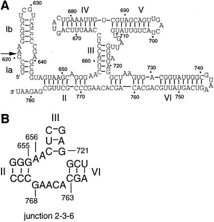
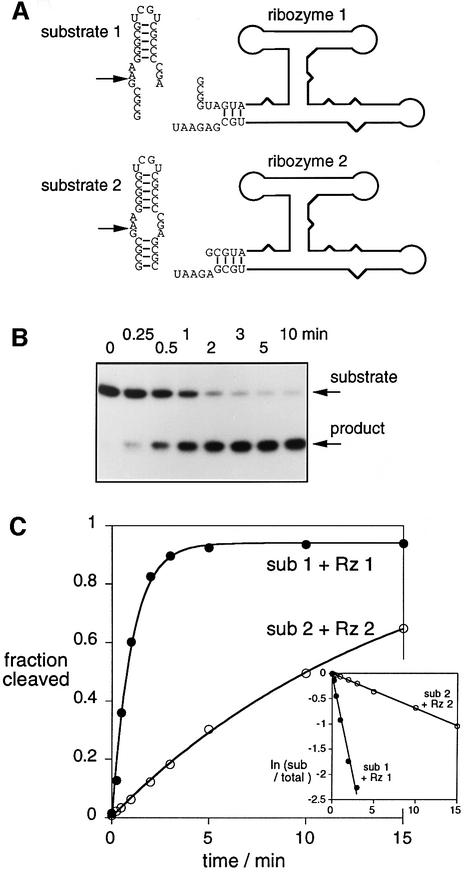
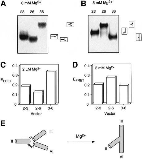
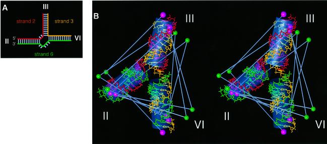
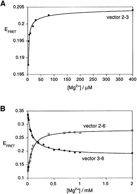
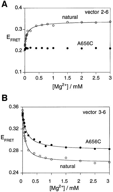
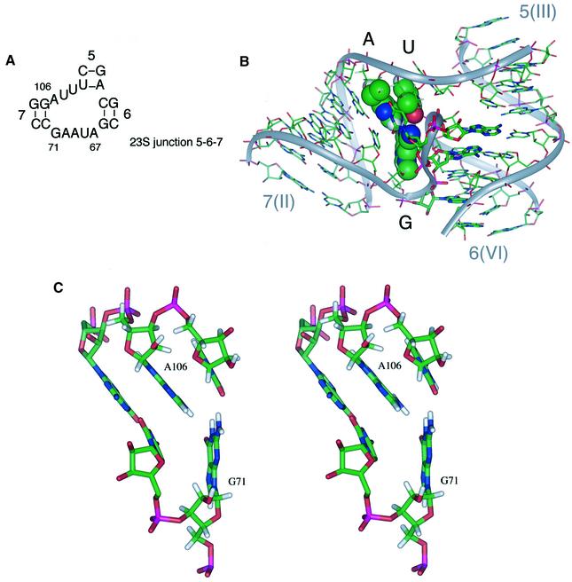
The VS nucleolytic ribozyme has a core comprising five helices organized by two three-way junctions. The ribozyme can act in trans on a hairpin-loop substrate, with which it interacts via tertiary contacts. We have determined that one of the junctions (2-3-6) undergoes two-stage ion-dependent folding into a stable conformation, and have determined the global structure of the folded junction using long-range distance restraints derived from fluorescence resonance energy transfer. A number of sequence variants in the junction are severely impaired in ribozyme cleavage, and there is good correlation between changes in activity and alteration in the folding of junction 2-3-6. These studies point to a special importance of G and A nucleotides immediately adjacent to helix II, and comparison with a similar junction of known structure indicates that this could adopt a guanine-wedge structure. We propose that the 2-3-6 junction organizes important aspects of the structure of the ribozyme to facilitate productive association with the substrate, and suggest that this results in an interaction between the substrate and the A730 loop to create the active complex.


Figures

None
Fig. 1. The sequence and secondary structure of the VS ribozyme. (A) The secondary structure comprises six helical sections, which can be considered as substrate (helix I, subdivided as Ia and Ib) and ribozyme (helices II–VI) (Beattie et al., 1995). Ribozyme cleavage occurs at the position indicated by the arrow. The conventional numbering of the nucleotides is indicated on the figure, as used throughout the text. (B) Junction 2-3-6 of the ribozyme, showing the numbering of selected nucleotides.
None
Fig. 2. Cleavage activity of the VS ribozyme in trans. (A) The RNA has been divided into substrate and ribozyme in two ways, as indicated in this schematic. In substrate 2, helix Ia comprises four intramolecular base pairs, whereas this helix is only generated in substrate 1 by intermolecular base pairing with ribozyme 1. (B) Cleavage of radioactive 5′-32P-labelled substrate 1 by ribozyme 1 as a function of time. The substrate and 5 nt product were separated by gel electrophoresis, and an autoradiograph of the gel is shown. The reaction timepoints are written above the gel. (C) Plotted reaction progress for the cleavage of substrate (sub) 1 by ribozyme (Rz) 1 (closed circles) and substrate 2 by ribozyme 2 (open circles). Single-exponential fits are shown by the lines. The insert shows plots of ln(fraction of uncleaved substrate) versus time for the same data.
None
Fig. 3. The global structure of the VS junction 2-3-6 studied by comparative gel electrophoresis and fluorescence resonance energy transfer (FRET). (A and B) Comparative gel electrophoresis. Three-way junctions were generated from strands in which the central 16 nt (plus the unpaired bases) were RNA and the remaining 32 nt at each end were DNA. The three possible species having two long arms of 40 bp and one shorter arm of 10 bp were created, and named according to the two long arms (e.g. species 23 has long II and III arms). The three long–short species were electrophoresed in a polyacrylamide gel in the presence of the indicated metal ion concentrations. The junctions were created from strands that were radioactively 5′-32P-labelled, and the different junction species were revealed by phosphorimaging of dried gels. The experiment was repeated in the presence of 0 (A) and 5 mM (B) magnesium ions. It is clear that the global structure is dependent on the prevailing metal ion concentration. The inter pretations of the mobility patterns are indicated to the right of the phosphorimages, corresponding to the global structures illustrated in (E). (C and D) FRET analysis. Data for species with arms of 12 bp are shown here. They are named according to the arms bearing fluorescein and Cy3, in that order. The data are presented as histograms of the efficiency of FRET (EFRET) for three end-to-end vectors, measured in the presence of 2 µM (C) or 2 mM (D) magnesium ions. (E) Global structure of the VS 2-3-6 junction in the presence and absence of magnesium ions, consistent with a qualitative interpretation of the experimental data.
None
Fig. 4. The global structure of the VS junction 2-3-6 folded in the presence of 2 mM magnesium ions, based on distance information derived from FRET measurements. (A) Schematic to illustrate the junctions used in the FRET analysis. The junctions comprise three strands, termed strands 2, 3 and 6 (taken from the arm containing the 5′ end of the strand). The colouring corresponds to that used in the structure shown in (B). (B) Stereographic representation of the low-violation structure for the junction in high magnesium ion concentration. Standard A-form helical sections are shown using a stick representation. The three strands are differentiated by colour; strand 2 is red, strand 3 is yellow and strand 6 is green. The positions of the fluorophores are indicated by single solid spheres (fluorescein, green; Cy3, pink). Grey bars indicate the FRET vectors used in the calculations. Overall helix orientations are indicated by semi-transparent grey cylinders.
None
Fig. 5. Folding of the VS junction 2-3-6 as a function of magnesium ion concentration. Conformational transitions were studied by the change of EFRET for the three end-to-end vectors: (A) 2-3 (closed circles), (B) 2-6 (open squares) and 3-6 (closed circles). Vector 2-3 undergoes a shortening at a magnesium ion concentration lower than that inducing the shortening and lengthening of the 2-6 and 3-6 vectors, respectively. The experimental data were fitted (lines) in each case by regression to a simple two-state model where the binding of metal ions to the RNA induces a structural change.
None
Fig. 6. Perturbed folding of a sequence variant of the VS junction 2-3-6. The folding of variant A656C was studied by FRET efficiency as a function of magnesium ion concentration. For these experiments, species with arm lengths of 11 bp were employed. Plots of EFRET as a function of magnesium ion concentration are shown for the natural and variant junctions. (A) Vector 2-6. Note that the FRET efficiency of the A656C variant (closed circles) is virtually unchanged over the complete range of ion concentration. This is markedly different from the behaviour of natural sequence junction (open circles). The data for the natural sequence junction have been fitted (line) to the two-state binding model as before. (B) Vector 3-6. This vector exhibits an ion-dependent change in FRET efficiency for the A656C variant (closed circles), but the eventual plateau value is higher than that of the natural sequence (open circles). Both sets of data have been fitted (lines) to the two-state binding model.
None
Fig. 7. The structure of the ribosomal RNA junction and the pivotal role of the guanine wedge. (A) The sequence of a junction occurring near the 5′ end of 23S rRNA, whose sequence is similar to that to the VS ribozyme. The helices have been sequentially numbered from the 5′-terminus of 23S rRNA, and the nucleotide numbering corresponds to that of Haloarcula marismortui. Positions 71 and 106 correspond to G75 and A111, respectively, in E.coli. The RNA sequences of both E.coli and H.marismortui conform to this consensus. In cases where A106 is mutated, it is always replaced by guanine. (B) The structure of the three-way junction from 23S ribosomal RNA (Ban et al., 2000) consisting of the intersection of helices 7, 5 and 6 (the equivalent helices in the VS ribozyme are II, III and VI). The bases G71, A106 and U107, forming the guanine-wedge feature, are shown in space-filling representation. (C) A stereographic representation of the guanine-wedge residues and the associated first base pair of helix 7. This representation shows clearly the proposed hydrogen-bond interactions between G71-O6 and A106-HN6, G71-HN2 and U107-O2. The angle formed between the bases G71 and A106 appears to be directly related to the angle subtended between helices 7 and 6.

Similar articles

Cited by

References

    1. Andersen A.A. and Collins,R.A. (2000) Rearrangement of a stable RNA secondary structure during VS ribozyme catalysis. Mol. Cell, 5, 469–478. - PubMed
    1. Ban N., Nissen,P., Hansen,J., Moore,P.B. and Steitz,T.A. (2000) The complete atomic structure of the large ribosomal subunit at 2.4 Å resolution. Science, 289, 905–920. - PubMed
    1. Bassi G., Møllegaard,N.E., Murchie,A.I.H., von Kitzing,E. and Lilley,D.M.J. (1995) Ionic interactions and the global conformations of the hammerhead ribozyme. Nature Struct. Biol., 2, 45–55. - PubMed
    1. Bassi G.S., Murchie,A.I.H. and Lilley,D.M.J. (1996) The ion-induced folding of the hammerhead ribozyme: core sequence changes that perturb folding into the active conformation. RNA, 2, 756–768. - PMC - PubMed
    1. Bassi G.S., Murchie,A.I.H., Walter,F., Clegg,R.M. and Lilley,D.M.J. (1997) Ion-induced folding of the hammerhead ribozyme: a fluorescence resonance energy transfer study. EMBO J., 16, 7481–7489. - PMC - PubMed

Publication types