. 2001 Mar 15;20(6):1439-48.
doi: 10.1093/emboj/20.6.1439.

5' cloverleaf in poliovirus RNA is a cis-acting replication element required for negative-strand synthesis

Affiliations

5' cloverleaf in poliovirus RNA is a cis-acting replication element required for negative-strand synthesis

D J Barton et al. EMBO J. .

Abstract

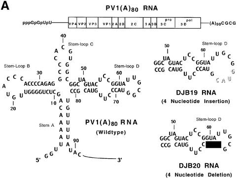
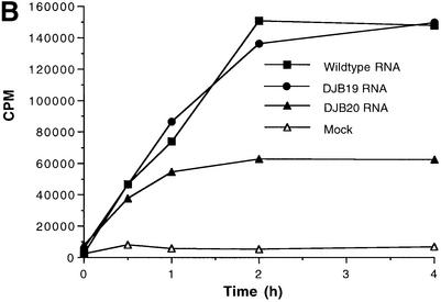
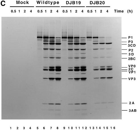
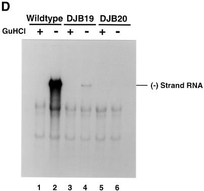
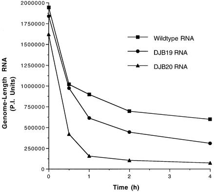
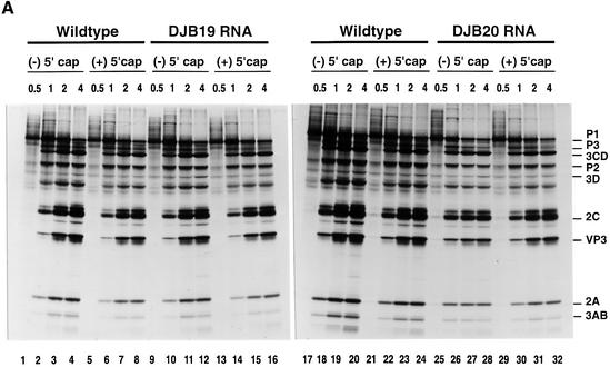
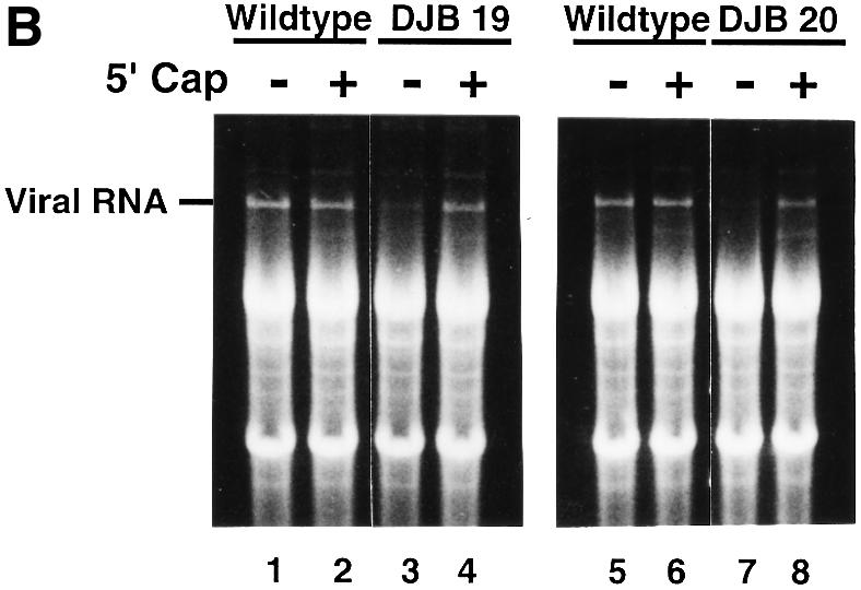
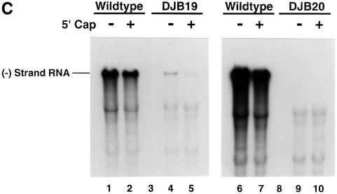
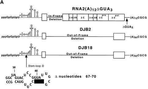
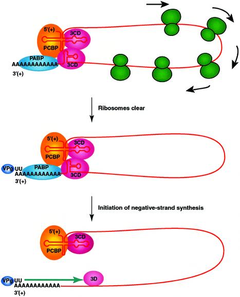
A cloverleaf structure at the 5' terminus of poliovirus RNA binds viral and cellular proteins. To examine the role of the cloverleaf in poliovirus replication, we determined how cloverleaf mutations affected the stability, translation and replication of poliovirus RNA in HeLa S10 translation-replication reactions. Mutations within the cloverleaf destabilized viral RNA in these reactions. Adding a 5' 7-methyl guanosine cap fully restored the stability of the mutant RNAs and had no effect on their translation. These results indicate that the 5' cloverleaf normally protects uncapped poliovirus RNA from rapid degradation by cellular nucleases. Preinitiation RNA replication complexes formed with the capped mutant RNAs were used to measure negative-strand synthesis. Although the mutant RNAs were stable and functional mRNAs, they were not active templates for negative-strand RNA synthesis. Therefore, the 5' cloverleaf is a multifunctional cis-acting replication element required for the initiation of negative-strand RNA synthesis. We propose a replication model in which the 5' and 3' ends of viral RNA interact to form a circular ribonucleoprotein complex that regulates the stability, translation and replication of poliovirus RNA.


Figures

None
Fig. 1. Effect of 5′ cloverleaf mutations on viral protein synthesis and negative-strand RNA synthesis. (A) Diagram of poliovirus transcript RNA, T7-PV1(A)80 RNA, and 5′ cloverleaf structure. The wild-type cloverleaf structure, the GUAC insertion in stem–loop D in DJB19 RNA and the GUAC deletion (black box) in stem–loop D in DJB20 RNA are shown. (B) Viral protein synthesis was measured in HeLa S10 in vitro translation–replication reactions containing either T7-PV1(A)80 RNA (wild type), DJB19 RNA or DJB20 RNA. The translation reactions contained [35S]methionine (1.2 mCi/ml) and 50 µg/ml RNA as indicated. At the indicated times, 1 µl samples were removed from each reaction. The labeled viral proteins were precipitated in 5% trichloroacetic acid, collected on filters and quantitated by scintillation counting. The amount of labeled protein synthesized in each reaction was plotted as a function of reaction time. (C) SDS–PAGE analysis of the labeled viral proteins. At the indicated times, 4 µl samples were removed from each translation reaction. The labeled viral proteins were solubilized in 50 µl of SDS sample buffer, denatured at 100°C for 3 min and 20 µl portions were separated by electrophoresis in an SDS–9–18% polyacrylamide gel. The gel was fixed and fluorographed. (D) Negative-strand RNA synthesis was measured using preinitiation RNA replication complexes isolated from HeLa S10 translation–RNA replication reactions containing guanidine HCL and each of the indicated RNAs using the procedures described in Materials and methods. Reactions containing the preinitiation RNA replication complexes were incubated at 37°C for 32 min and 32P-labeled negative-strand RNA was fractionated by CH3HgOH–agarose gel electrophoresis. The position of poliovirus negative-strand RNA is indicated.
None
Fig. 2. Mutations in 5′ cloverleaf structure decrease the stability of poliovirus RNA. Four micrograms of 32P-labeled T7-PV1(A)80 RNA, DJB19 RNA or DJB20 RNA (∼580 000 c.p.m./µg) were added to 50 µl of HeLa S10 translation–RNA replication reactions containing 2 mM guanidine HCl. The reactions were incubated at 34°C and 7 µl samples were removed at the indicated times and added to 300 µl of 0.5% SDS buffer. The labeled RNA samples were phenol extracted, ethanol precipitated and analyzed by electrophoresis in a CH3HgOH–agarose gel. The amount of full-length labeled viral RNA detected at each time point was quantitated using PhosphorImager (PI) analysis. Arbitrary PI units of full-length viral RNA were plotted versus incubation time.
None
Fig. 3. Capped DJB19 and DJB20 RNAs are stable and translate at wild-type or slightly reduced levels but are not functional templates for negative-strand synthesis. (A) Labeled viral protein synthesis was measured as described in Figure 1 using HeLa S10 translation–RNA replication reactions that contained T7-PV1(A)80 RNA (wild type), DJB19 RNA or DJB20 RNA (50 µg/ml) with or without a 5′ cap as indicated. Labeled viral proteins synthesized at each time point were analyzed by SDS–PAGE as described in Figure 1C. (B and C) The stability of the input RNAs and negative-strand RNA synthesis was measured in reactions containing preinitiation RNA replication complexes that were isolated from HeLa S10 translation–RNA replications containing guanidine HCl and the indicated viral RNAs. The reactions were incubated at 37°C for 30 min and the total RNA and the labeled product RNAs were analyzed by CH3HgOH–agarose gel electrophoresis as described in Materials and methods. (B) UV light visualization of total RNA within the gels after being stained with ethidium bromide is shown. The position of the input viral RNA in the gel is indicated. (C32P-labeled negative-strand RNA detected by autoradiography of the dried gels. The position of 32P-labeled poliovirus negative-strand RNA is marked.
None
Fig. 4. Complementation analysis of DJB2 and DJB18 RNA replication in vitro. (A) Diagram of RNA2(A)12ΔGUA3, the helper RNA used in this experiment. The co-translation of this helper RNA provided the viral replication proteins in trans in reactions containing viral transcript RNAs that contained a deletion in the coding sequence for the viral replication proteins. DJB2 RNA does not encode any of the viral replication proteins but was shown in a separate study in our laboratory to contain all of the cis-active replication elements required to act as a template for negative-strand RNA synthesis. DJB18 RNA is identical to DJB2 RNA except for the deletion of poliovirus nucleotides 67–70 in the 5′ cloverleaf. (B) HeLa S10 translation–RNA replication reactions (100 µl) that contained 2 mM guanidine HCl and the indicated RNA (±) 5′ cap were incubated for 4 h at 34°C. The helper RNA was added at a 2:1 molar ratio relative to DJB2 RNA or DJB18 RNA, and the total RNA concentration was maintained at 100 µg/ml in each reaction. Negative-strand RNA synthesis was measured in preinitiation RNA replication complexes that were isolated from these reactions as described in Materials and methods. Reactions containing the preinitiation RNA replication complexes were incubated at 37°C for 25 min, and labeled negative-strand synthesis was analyzed by CH3HgOH–agarose gel electrophoresis. The mobility of DJB2 RNA, DJB18 RNA and the helper RNAs is shown. Control experiments showed that equivalent amounts of the viral replication proteins were synthesized in each reaction that contained the helper RNA (data not shown).
None
Fig. 5. Model of circular RNP complex that is used to initiate negative-strand RNA synthesis. Viral proteins 3CD and VPg, poly(A) binding protein (PABP) and poly(rC) binding protein (PCBP) interact with each other and the ends of the viral RNA to form a circular RNP complex. The inhibition of translation initiation allows for the clearance of the viral RNA over time by the elongation of translating ribosomes to the 3′ end of the coding sequence (top). Once the template RNA is cleared of translating ribosomes, VPg-pUpU (or VPg) associates with the 3′ end of the viral RNA template and completes the formation of the circular preinitiation RNA replication complex (middle). Negative-strand synthesis initiates by the elongation of the VPg-primer by the viral polymerase, 3Dpol (bottom). Additional viral and cellular proteins, proteolytic processing events and cellular membranes are also required for viral RNA replication but for clarity are not depicted in this model. See text for additional details.

References

    1. Ambros V. and Baltimore,D. (1980) Purification and properties of a HeLa cell enzyme able to remove the 5′-terminal protein from poliovirus RNA. J. Biol. Chem., 255, 6739–6744. - PubMed
    1. Ambros V., Pettersson,R.F. and Baltimore,D. (1978) An enzymatic activity in uninfected cells that cleaves the linkage between poliovirion RNA and the 5′ terminal protein. Cell, 15, 1439–1446. - PubMed
    1. Andino R., Rieckhof,G.E. and Baltimore,D. (1990a) A functional ribonucleoprotein complex forms around the 5′ end of poliovirus RNA. Cell, 63, 369–380. - PubMed
    1. Andino R., Rieckhof,G.E., Trono,D. and Baltimore,D. (1990b) Substitutions in the protease (3Cpro) gene of poliovirus can suppress a mutation in the 5′ noncoding region. J. Virol., 64, 607–612. - PMC - PubMed
    1. Andino R., Rieckhof,G.E., Achacoso,P.L. and Baltimore,D. (1993) Poliovirus RNA synthesis utilizes an RNP complex formed around the 5′-end of viral RNA. EMBO J., 12, 3587–3598. - PMC - PubMed

Publication types