. 2001 Mar 15;20(6):1462-8.
doi: 10.1093/emboj/20.6.1462.

The DnaB.DnaC complex: a structure based on dimers assembled around an occluded channel

Affiliations

The DnaB.DnaC complex: a structure based on dimers assembled around an occluded channel

M Bárcena et al. EMBO J. .

Abstract

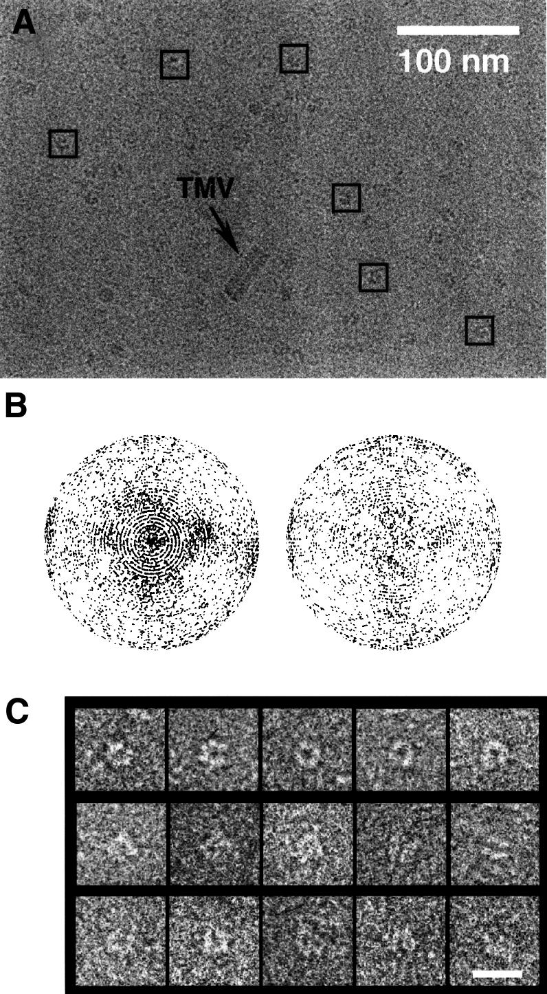
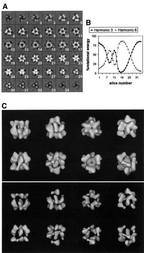
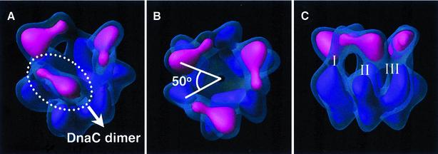
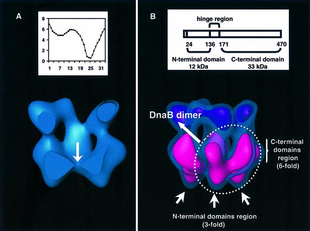
Replicative helicases are motor proteins that unwind DNA at replication forks. Escherichia coli DnaB is the best characterized member of this family of enzymes. We present the 26 A resolution three-dimensional structure of the DnaB hexamer in complex with its loading partner, DnaC, obtained from cryo-electron microscopy. Analysis of the volume brings insight into the elaborate way the two proteins interact, and provides a structural basis for control of the symmetry state and inactivation of the helicase by DnaC. The complex is arranged on the basis of interactions among DnaC and DnaB dimers. DnaC monomers are observed for the first time to arrange as three dumb-bell-shaped dimers that interlock into one of the faces of the helicase. This could be responsible for the freezing of DnaB in a C(3) architecture by its loading partner. The central channel of the helicase is almost occluded near the end opposite to DnaC, such that even single-stranded DNA could not pass through. We propose that the DnaB N-terminal domain is located at this face.


Figures

None
Fig. 1. Cryo-EM of the DnaB·DnaC complex. (A) Electron micrograph of a vitrified preparation of E.coli DnaB·DnaC imaged at 0° tilt. Some particles selected from this micrograph are indicated by squares. (B) Angular distribution of the projection images from untilted (left) and tilted (right) micrographs. Each projection image is represented by a point in a sphere, viewed in the direction of the polar axis. Pure top views are at the pole of the hemisphere, whereas pure side views are on the equator. The longitude of each point represents the azimuthal angle of the projection image. (C) Galleries of top views (first row), side views (second row) and intermediate views (third row). The bar represents 20 nm.
None
Fig. 2. DnaB·DnaC complex at 26 Å resolution. (A) Slices through the reconstructed volume. Significant slices extend from 1 to 34. (B) Variations in symmetry along the rotational axis of the volume. The plots represent the relative energy of the 3- and 6-fold components of the rotational power spectra of successive slices. (C) Surface renderings of the volume at a threshold accounting for 100% (top) and 40% (bottom) of the expected mass. The volume was symmetrized and filtered to its resolution.
None
Fig. 3. DnaC in the complex. Surface renderings in semi-transparent blue account for 100% of the expected mass and those in opaque colours account for 40%. In the latter, the regions considered to be DnaC are depicted in pink, and the three DnaC dimers become segmented apart (A). There exists a relative twist among DnaC and DnaB dimers of ∼50° (measured as the relative rotation between the higher density areas of a DnaC dimer and a neighbouring DnaB dimer) (B). Three different contact areas (I, II and III) between each DnaC dimer and DnaB can be clearly distinguished (C).
None
Fig. 4. DnaB in the complex. (A) Surface rendering representation of the DnaB·DnaC complex (100% of the mass) cut by a central plane through the length of the central channel. The arrow points towards the region where the channel narrows sharply. (Inset) Variations in the width of the central channel; the diameter of the channel (in nm) is plotted versus the slice number (Figure 2A). (B) Assignment of DnaB domains within the volume. The domain organization of the DnaB monomer is summarized in the scheme (inset), where numbers refer to residue positions in the primary structure. The reconstruction is presented at two different threshold values as in Figure 3. In this case, in the surface rendering that accounts for 40% of the mass, the region considered to be DnaB is highlighted in pink.

Similar articles

Cited by

References

    1. Baker T.A. and Bell,S.P. (1998) Polymerases and the replisome: machines within machines. Cell, 92, 295–305. - PubMed
    1. Baker T.A., Sekimizu,K., Funnell,B.E. and Kornberg,A. (1986) Extensive unwinding of the plasmid template during staged enzymatic initiation of DNA replication from the origin of the Escherichia coli chromosome. Cell, 45, 53–64. - PubMed
    1. Bárcena M. (2000) Análisis estructural del polimorfismo cuaternario en las helicasas replicativas y del complejo DnaB⋅DnaC de Escherichia coli. PhD thesis, Universidad Autónoma de Madrid, Madrid, Spain.
    1. Biswas S.B., Chen,P.-H. and Biswas,E.E. (1994) Structure and function of Escherichia coli DnaB protein: role of the N-terminal domain in helicase activity. Biochemistry, 33, 11307–11314. - PubMed
    1. Chang P. and Marians,K.J. (2000) Identification of a region of Escherichia coli DnaB required for functional interaction with DnaG at the replication fork. J. Biol. Chem., 275, 26187–26195. - PubMed

Publication types