Review
. 2000 Dec;1(3):171-81.
doi: 10.1038/35042039.

The establishment of Spemann's organizer and patterning of the vertebrate embryo

Affiliations
Review

The establishment of Spemann's organizer and patterning of the vertebrate embryo

E M De Robertis et al. Nat Rev Genet. 2000 Dec.

Abstract

Molecular studies have begun to unravel the sequential cell-cell signalling events that establish the dorsal-ventral, or 'back-to-belly', axis of vertebrate animals. In Xenopus and zebrafish, these events start with the movement of membrane vesicles associated with dorsal determinants. This mediates the induction of mesoderm by generating gradients of growth factors. Dorsal mesoderm then becomes a signalling centre, the Spemann's organizer, which secretes several antagonists of growth-factor signalling. Recent studies have led to new models for the regulation of cell-cell signalling during development, which may also apply to the homeostasis of adult tissues.


Figures

Figure 1
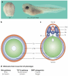
Figure 1. The anatomy of Xenopus development
The ovarian oocyte is radially symmetrical and is divided into an animal and a vegetal domain. One hour after fertilization, an unpigmented dorsal crescent is formed in the fertilized egg opposite the sperm entry point. As the embryo rapidly divides into smaller and smaller cells, without intervening growth (cleavage), a cavity called the blastocoel is formed, which defines the blastula stage. By the late blastula stage (9 h of development), the three germ layers become defined. The ectoderm, or animal cap, forms the roof of the blastocoel. The mesoderm is formed in a ring of cells in the marginal zone, located between the ectoderm and endoderm. At the gastrula stage (10 h), involution of the mesoderm towards the inside of the embryo starts at the dorsal blastopore lip. The morphogenetic movements of gastrulation lead to the formation of the vertebrate body plan, patterning the ectoderm, mesoderm and endoderm. At the neurula stage (14 h), the neural plate, or future central nervous system (CNS), becomes visible in dorsal ectoderm. By the tailbud stage (24-42 h), a larva with a neural tube located between the epidermis and the notochord has formed. The blastopore gives rise to the anus, and the mouth is generated by secondary perforation.
Figure 2
Figure 2. The ultraviolet (UV) phenotype of irradiated embryos can be rescued by many different molecules
a | Ultraviolet-irradiated embryos do not develop axial structures, and differentiate into a belly piece (left). Microinjection of synthetic mRNA for Chordin, an antagonist of bone morphogenetic protein (BMP) signalling, completely rescues the development of axial structures (right). b | The belly piece comprises epidermis (Ep), lateral plate mesoderm (LPM), endoderm (En) and blood (Bl), whereas c | unirradiated or ultraviolet-treated embryos injected with chordin mRNA differentiate dorsal tissues, such as central nervous system (CNS), somites (So), notochord (No), kidney (Ki) and hypochord (Hc). d | A puzzling variety of gene products can rescue the ultraviolet phenotype; the main thesis explored in this review is that these molecules participate in a sequential pathway of biochemical events leading to dorsal development.
Figure 3
Figure 3. Dorsal determinants and the transport of membrane vesicles to the dorsal side
a | After fertilization, parallel arrays of cortical microtubules extend from the centriole and the large aster at the sperm entry point towards the dorsal side (where the plus end lies), and transport small membrane vesicles from the vegetal towards the dorsal animal pole. The inset shows that the dorsal determinant vesicles are associated with Dishevelled (Dsh), a component of the Wnt signal transduction pathway. The Wnt receptor Frz7 is required for dorsal axis formation, but the Wnt molecules shown inside the vesicles are entirely hypothetical. Kinesins are molecular motors that can transport vesicles towards the plus ends of microtubules. β-Catenin is found in a large cytoplasmic complex that includes Axin, APC (adenomatous polyposis coli) and GSK-3 (glycogen synthase kinase 3). GSK-3 negatively regulates β-Catenin through phosphorylations that target β-Catenin for degradation by the proteasome. GSK-3 can be inhibited by treatment with lithium chloride (LiCl). On stimulation of the Wnt pathway, β-Catenin is stabilized on the dorsal side and can be found in the nucleus, where, together with TCF-3, it activates various target genes, including homeobox and Nodal-related genes (Xnrs). Further molecules that participate in this canonical Wnt signalling pathway are not shown in this simplified diagram. b | Irradiation of embryos with ultraviolet light disrupts cortical microtubules and prevents the transport of the membrane vesicles to the prospective dorsal side.
Figure 4
Figure 4. Two-step model of mesoderm induction in Xenopus
At the midblastula stage, higher β-Catenin levels on the dorsal side of the embryo, together with the vegetally located transcription factor VegT and the maternal TGF-β-family growth factor Vg1, generate a gradient of Nodal-related molecules expressed in the endoderm. In turn, this gradient induces the formation of overlying mesoderm: low doses of Nodal-related molecules (Xnrs) lead to the formation of ventral mesoderm, whereas high doses lead to the establishment of Spemann’s organizer. Nieuwkoop’s centre is the region of dorsal endoderm that induces organizer tissue. At the gastrula stage, the organizer secretes a cocktail of factors that refine the initial patterning. Note that β-Catenin is widely distributed on the dorsal side, including in derivatives of the three germ layers. (CNS, central nervous system.)
Figure 5
Figure 5. Spemann’s organizer is a source of secreted growth factor antagonists
The organizer secretes proteins that bind to different growth factors in the extracellular space and block signalling through their cognate receptors. Crescent, Frzb-1 and Dickkopf-1 are Wnt antagonists. Cerberus is a multivalent inhibitor that antagonizes Xwnt-8, Xnrs and BMPs. Chordin, Noggin and Follistatin bind to and inhibit bone morphogenetic proteins (BMPs). Lefty/Antivin blocks Nodal signalling through a different mechanism, by binding to the TGF-β/Nodal receptor and acting as a competitive inhibitor.
Figure 6
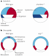
Figure 6. Genetics of chordin/sog in zebrafish and Drosophila
a | In zebrafish, loss-of-function mutations in chordino ventralize the mesoderm and the ectoderm. Organizer tissue and neuroectoderm are reduced, and ventral mesoderm and epidermis are expanded in chordino-/- embryos. b | In Drosophila, mutations in sog lead to dorsalized embryos. The neurogenic ectoderm is reduced, the amnioserosa is lost, and the amount of tissue allocated to dorsal ectoderm is expanded. The amnioserosa is the dorsal-most tissue of the fruitfly embryo and forms a thin layer of cells required for proper gastrulation. Formation of the amnioserosa requires the activity of Sog and dTsg. Dorsal-ventral polarity in arthropods has been inverted with respect to that of vertebrates in the course of evolution.
Figure 7
Figure 7. A molecular pathway involving Chordin, Xolloid and Twisted-gastrulation regulates the dorsal-ventral activity gradient of bone morphogenetic protein in Xenopus
a | Chordin binds bone morphogenetic protein (BMP) through cysteine-rich domains CR1 and CR3. After cleavage of Chordin by Xolloid (small arrows), the CR modules still bind BMP, but with tenfold reduced affinity. In principle, this lowered affinity might explain why Xolloid cleavage restores BMP signalling from inactive BMP-Chordin complexes. b | In the presence of Twisted-gastrulation (xTsg) protein, two new activities are observed. First, xTsg increases the binding of BMP to full-length Chordin, converting the Chordin-xTsg-BMP ternary complex into a better BMP antagonist. Second, after Xolloid cleavage, residual binding of BMP to CR modules is readily competed by xTsg. The binary xTsg-BMP complex does not interfere with BMP receptor binding at physiological concentrations, therefore eliminating inhibition by CR fragments and providing a permissive signal for BMP binding to its cognate receptors. The thicker arrows in b indicate the effects of xTsg on this biochemical pathway. Activated BMP receptor is phosphorylated (circled Ps), relaying the signal intracellularly.

References

    1. Spemann H. Vererbung und Entwicklungsmechanik. Naturwissenchaften. 1924;12:65–79.
    1. Hamburger V. The Heritage of Experimental Embryology: Hans Spemann and the Organizer. Oxford Univ. Press; Oxford: 1988. A magnificent account of the experiments that have shaped embryological thinking in the twentieth century.
    1. Brachet J. An old enigma: the gray crescent of amphibian eggs. Curr. Top. Dev. Biol. 1977;11:133–186. - PubMed
    1. Gerhart J, Doniach T, Stewart R. In: Gastrulation: Movements, Patterns, and Molecules. Keller R, Clark WH, Griffin F, editors. Plenum; New York: 1991. pp. 57–76.
    1. Black SD, Gerhart J. High frequency twinning of Xenopus laevis embryos from eggs centrifuged before first cleavage. Dev. Biol. 1986;116:228–240. - PubMed

Publication types