. 2001 Apr;69(4):2054-65.
doi: 10.1128/IAI.69.4.2054-2065.2001.

Identification of new genes involved in the virulence of Listeria monocytogenes by signature-tagged transposon mutagenesis

Affiliations

Identification of new genes involved in the virulence of Listeria monocytogenes by signature-tagged transposon mutagenesis

N Autret et al. Infect Immun. 2001 Apr.

Abstract

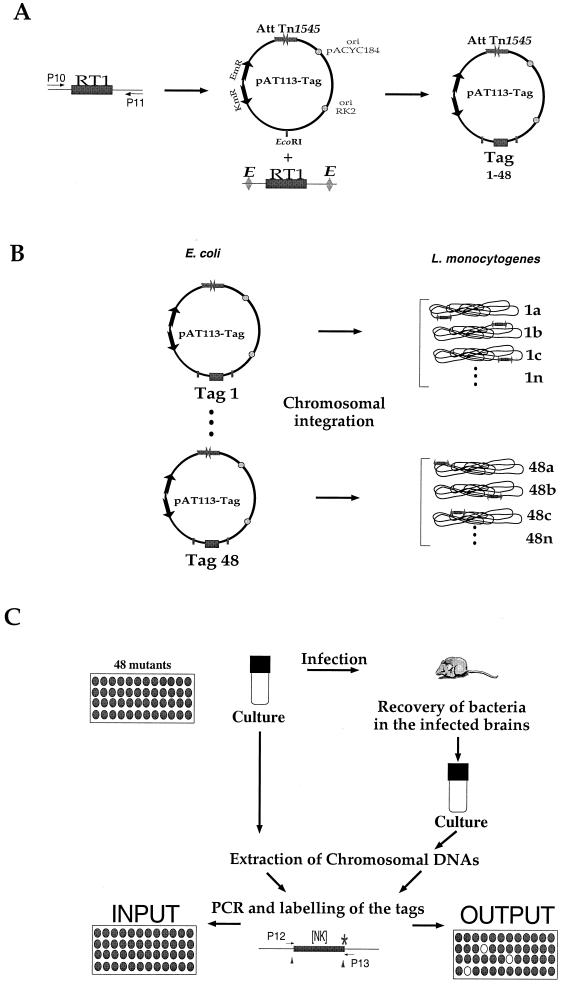
Listeria monocytogenes is a gram-positive, facultative intracellular pathogen that can cause severe food-born infections in humans and animals. We have adapted signature-tagged transposon mutagenesis to L. monocytogenes to identify new genes involved in virulence in the murine model of infection. We used transposon Tn1545 carried on the integrative vector pAT113. Forty-eight tagged transposons were constructed and used to generate banks of L. monocytogenes mutants. Pools of 48 mutants were assembled, taking one mutant from each bank, injected into mice, and screened for those affected in their multiplication in the brains of infected animals. From 2,000 mutants tested, 18 were attenuated in vivo. The insertions harbored by these mutants led to the identification of 10 distinct loci, 7 of which corresponded to previously unknown genes. The properties of four loci involving putative cell wall components were further studied in vitro and in vivo. The data suggested that these components are involved in bacterial invasion and multiplication in the brain.


Figures

FIG. 1
FIG. 1
Plasmids, primers, and screening strategy. (A) Construction of the tagged transposons. Schematic map of RT1 (P10 and P11 are the primers used for amplification the random oligonucleotide signature tag) and of the integrative plasmid pAT113. After digestion with EcoRI, PCR-amplified RT1 was introduced into the EcoRI site of pAT113 (tags 1 to 48 symbolize the different tags cloned). The 48 tagged pAT113 plasmids were constructed in E. coli. Abbreviations: AttTn1545, attachment site of Tn1545; ori (RK2 and pACYC184), origin of replication; EmR, resistance to ERY encoded by the erm gene, KmR, KAN restance encoded by the aphA-3 gene. (B) Generation of the banks of L. monocytogenes mutants. The 48 tagged pAT113 plasmids were introduced individually into L. monocytogenes to generate 48 banks of mutants (numbered 1 to 48); a, b, c, and n represent the different clones within each bank (i.e., carrying the same tag). (C) Screening strategy. The basic STM schema was adapted. Pools of 48 L. monocytogenes mutants were screened in vivo. Four days after inoculation, the brains of infected mice were recovered and used to inoculate bacterial cultures. The extracted chromosomal DNAs were then used for PCR amplification of the tags. [NK], the random portion of the tag that was amplified using primers P12 and P13.
FIG. 2
FIG. 2
Locus gtcA. (A) Schematic organization of the gtcA region. (Upper part) rpmE-rho region of B. subtilis. The rpmE is shaded in dark gray; the rho is shaded in light gray. (Middle part) rpmE-gtcA-rho region of L. monocytogenes serotype 4b. (Lower part) rpmE-csbB-gtcA-rho region of L. monocytogenes serotype 1/2a. (B) Sequence alignment of the GtcA from serotype 4b and 1/2a strains. Sequence alignment was performed using the CLUSTALw algorithm (www.infobiogen.fr/services/analyseq/cgi-bin/clustalw_in.pl). Similar residues are shaded, and identical residues are boxed (thick lines). The numbers refer to amino acid length.
FIG. 3
FIG. 3
Locus celR. (Upper part) Schematic organization of the celR locus in B. subtilis. The three domains comprised within the FruA polypeptide are indicated (IIA, IIB, and IIC). (Lower part) Schematic organization of the celR locus in L. monocytogenes. The three genes downstream of celR determine three ORFs that share 34, 44, and 52% identities, respectively, with the corresponding domains of the FruA protein of B. subtilis. The arrows indicate the approximate size and orientation of the different genes.
FIG. 4
FIG. 4
Southern blot of PstI-digested genomic DNA from the different mutant strains. A total of 5 μg of digested DNA was applied to 0.8% agarose gel, electrophoresed, blotted on nylon filter, and subjected to Southern hybridization. The 32P-labeled probe used was obtained after PCR amplification of a 0.7-kb fragment of pAT113. In the case of mutant YtgP, two identical clones were tested (E26 and H′49, in the 6th and last lanes, respectively). As a negative control (−), we used chromosomal DNA from EGD (wild type). The positions of the molecular weight markers are indicated in kilobases preceded by arrows on the right of the figure.
FIG. 5
FIG. 5
RT-PCR transcriptional analysis. Expression of the 10 loci identified was examined in EGD grown in BHI medium. Appropriate pairs of primers internal to each gene were used for RT-PCR. Amplified DNA fragments were detected after electrophoresis in 1% agarose gels by ethidium bromide staining. One microgram of molecular weight DNA ladder (MW; Gibco-BRL) was loaded at both sides of the gel. The fragments were arbitrarily loaded from the largest to the smallest (left to right).
FIG. 6
FIG. 6
In vivo survival of the attenuated strains. The kinetics of bacterial growth was monitored in the organs of mice infected with the different Tn1545 insertion mutants and compared with EGD as a positive control. Mice were inoculated with 106 bacteria (indicated by an arrow to the left of the ordinate). Bacterial survival was monitored in the spleen (A), liver (B), and brain (C) over a 6-day period. Symbols: ■, EGD; □, ORF626; ●, PbpX; ▴, GtcA; ○, YtgP. The point of death is indicated by a cross symbol.
FIG. 7
FIG. 7
Invasiveness of the attenuated strains. Invasiveness was evaluated in three different types of cell lines (A, Caco2 cells; B, Vero cells; and C, HepG-2 cells). Cell monolayers were incubated for 1 h at 37°C with approximately 200 bacteria per cell. After being washed, the cells were reincubated for 8 h in fresh culture medium containing gentamicin (10 mg liter−1). At intervals, the cells were washed again and lysed, and viable bacteria were counted on BHI-ERY plates. Datum points and error bars (indicated only for the wild-type strain for clarity) represent the mean and standard deviation of the number of bacteria per well. Three different assays were performed. Symbols: ■, EGD; □, ORF626; ●, PbpX; ▴, GtcA; ○, YtgP.

References

    1. Altschul S F, Madden T L, Schaffer A A, Zhang J, Zhang Z, Miller W, Lipman D J. Gapped BLAST and PSI-BLAST: a new generation of protein database search programs. Nucleic Acids Res. 1997;25:3389–3402. - PMC - PubMed
    1. Ausubel F M, Brent R, Kingston R E, Moore D D, Smith J A, Seidman J G, Struhl K. Current protocols in molecular biology. New York, N.Y: Wiley Interscience; 1990.
    1. Badger J L, Wass C A, Weissman S J, Kim K S. Application of signature-tagged mutagenesis for identification of Escherichia coli K1 genes that contribute to invasion of human brain microvascular endothelial cells. Infect Immun. 2000;68:5056–5061. - PMC - PubMed
    1. Berche P. Bacteremia is required for invasion of the murine central nervous system by Listeria monocytogenes. Microb Pathog. 1995;18:323–336. - PubMed
    1. Berche P, Gaillard J L, Sansonetti P J. Intracellular growth of Listeria monocytogenes as a prerequisite for in vivo induction of T cell-mediated immunity. J Immunol. 1987;138:2266–2271. - PubMed

Publication types

LinkOut - more resources