. 2001 Apr;69(4):2549-57.
doi: 10.1128/IAI.69.4.2549-2557.2001.

Haemophilus ducreyi associates with phagocytes, collagen, and fibrin and remains extracellular throughout infection of human volunteers

Affiliations

Haemophilus ducreyi associates with phagocytes, collagen, and fibrin and remains extracellular throughout infection of human volunteers

M E Bauer et al. Infect Immun. 2001 Apr.

Abstract

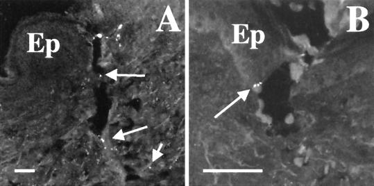
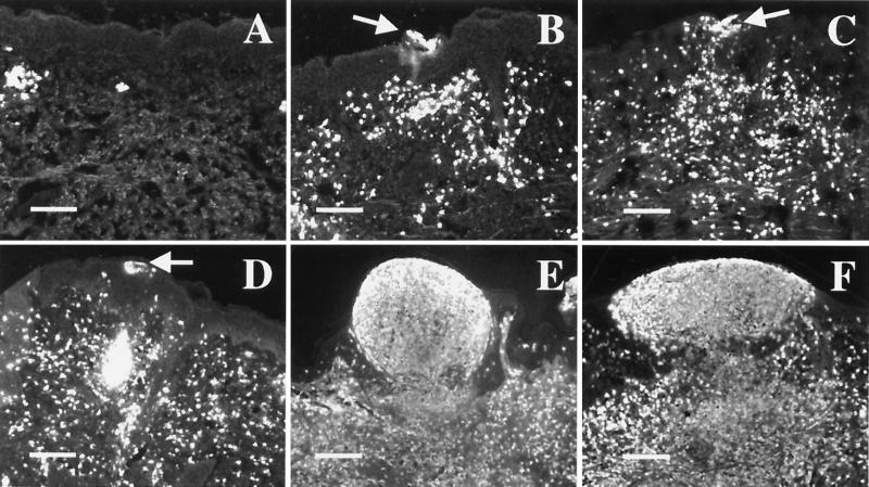
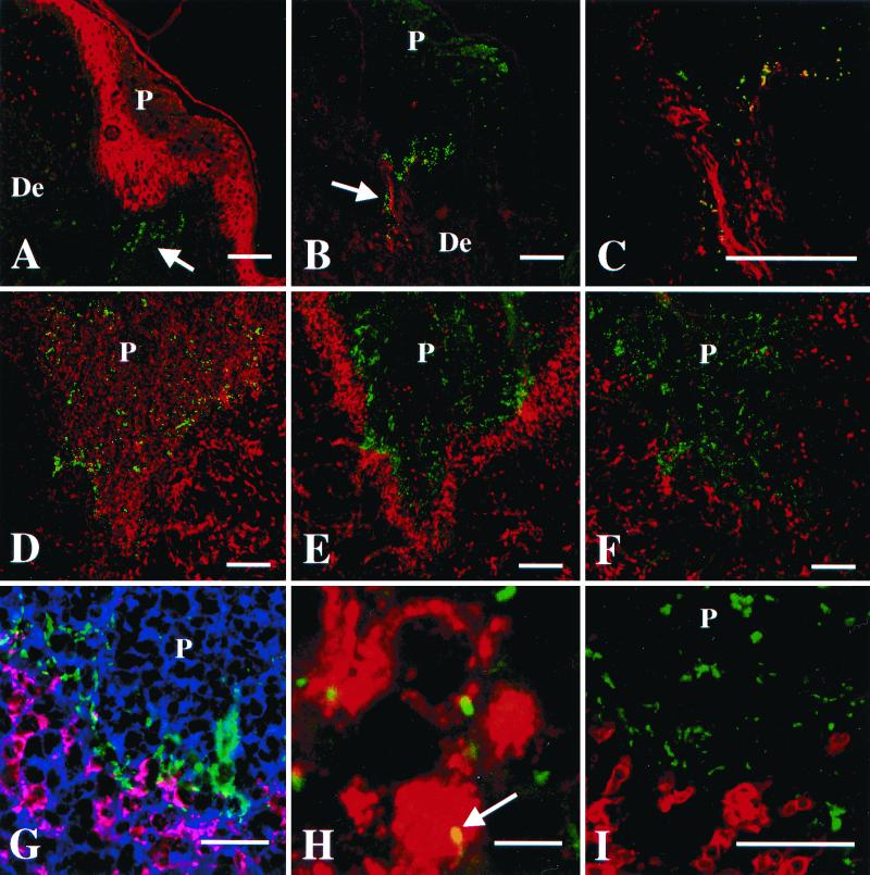
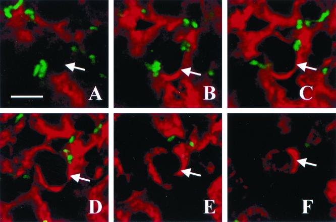
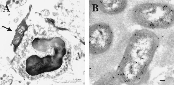
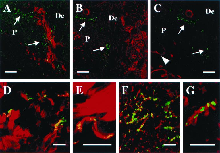
In a previous study, Haemophilus ducreyi was found in the pustule and dermis of samples obtained at the clinical end point in the human model of infection. To understand the kinetics of localization, we examined infected sites at 0, 24, and 48 h after inoculation and at the clinical end point. Immediately after inoculation, bacteria were found predominantly in the dermis but also in the epidermis. Few bacteria were detectable at 24 h; however, by 48 h, bacteria were readily seen in the pustule and dermis. H. ducreyi was associated with polymorphonuclear leukocytes and macrophages in the pustule and at its base, but was not associated with T cells, Langerhans' cells, or fibroblasts. H. ducreyi colocalized with collagen and fibrin but not laminin or fibronectin. Association with phagocytes, collagen, and fibrin was seen as early as 48 h and persisted at the pustular stage of disease. Optical sectioning by confocal microscopy and transmission electron microscopy both failed to demonstrate intracellular H. ducreyi. These data identify collagen and fibrin as potentially important targets of adherence in vivo and strongly suggest that H. ducreyi remains extracellular throughout infection and survives by resisting phagocytic killing in vivo.


Figures

FIG. 1
FIG. 1
Deposition of H. ducreyi in skin. Sections from tissue biopsied immediately postinoculation were stained with polyclonal anti-H. ducreyi antiserum and MAb BB11. (A) Puncture wound made by inoculation device. Arrows indicate bacteria along puncture wound. (B) Bacteria (arrow) at the epidermis with keratinocytes. Ep, epidermis. Bars, 50 μm.
FIG. 2
FIG. 2
Kinetics of lesion formation in the human model. Sections were stained with anti-PMN elastase. Panels represent tissue harvested immediately after inoculation (A), an erythematous macule at 24 h (B), papules at 24 h (C and D), a papule at 48 h (E), and a pustule at 48 h (F). Arrows indicate intraepidermal micropustules in 24-h samples. Bars, 150 μm.
FIG. 3
FIG. 3
Localization of H. ducreyi in tissues from papular and pustular stages of disease. P, pustule or micropustule. De, dermis. Scale bars represent 100 μm in panels A through F, 50 μm in panels G and I, and 5 μm in panel H. (A) Papule at 48 h doubly stained with polyclonal anti-H. ducreyi antiserum (green) and anticytokeratin MAbs (red). Arrow indicates bacteria (green) in dermis below micropustule. (B) A 48-h papule doubly stained with polyclonal anti-type I collagen (red) and the anti-H. ducreyi MAb BB11 (green). Arrow indicates bacteria colocalizing with collagen (yellow). (C) Section stained as in panel B showing colocalization (yellow) of H. ducreyi and collagen. (D through F) Low-power view of consecutive sections from one pustule stained with polyclonal anti-H. ducreyi antiserum (green) and MAb recognizing PMN elastase (D), CD68 (E), or CD3 (F) (red). (G) Higher-power view of the base of a pustule. Section was stained with polyclonal anti-H. ducreyi antiserum (green), anti-CD68 (red), and the lectin LCA, which stains plasma membranes (blue). Colocalization of anti-CD68 (red) and LCA (blue) on the macrophage cell surface is represented in pink. Note the bacteria between the macrophages and the PMNs of the pustule. (H) High-power view of section stained with polyclonal anti-H. ducreyi antiserum (green) and anti-CD68 (red). Arrow indicates bacteria colocalizing with a macrophage (yellow). (I) Base of a pustule stained with polyclonal anti-H. ducreyi antiserum (green) and anti-CD3 (red). Note that the bacteria do not colocalize with the T cells.
FIG. 4
FIG. 4
Optical sectioning through a pustule. Panels A through F represent, in order, images taken in 1-μm steps through a section stained with polyclonal anti-H. ducreyi antiserum (green) and anti-PMN elastase MAb (red). The arrow in each panel points to one edge of the PMN. Note the bacteria on the outside but not within the PMN. Bar, 5 μm.
FIG. 5
FIG. 5
Transmission electron micrographs of H. ducreyi in a pustule. (A) H. ducreyi at the base of the pustule surrounded by fibrin (arrow) and adjacent to a PMN. Bar, 1 μm. (B) Immunogold labeling of H. ducreyi with polyclonal anti-H. ducreyi antiserum and gold-conjugated secondary Ab. Bar, 0.1 μm.
FIG. 6
FIG. 6
H. ducreyi association with ECM proteins at the pustular stage. (A through C) Low-power images at the pustule base of consecutive tissue sections stained for H. ducreyi (green) and for collagen (A), fibronectin (B), or laminin (C) (red). Arrows indicate bacteria at the pustule base. Arrowhead in panel C indicates remains of the basement membrane. Bars, 100 μm. (D) Section stained with polyclonal anti-type I collagen (red) and MAb BB11 (green). Note the colocalizing signals between collagen and H. ducreyi (yellow). Bar, 25 μm. (E) Section stained as in panel D. Bar, 25 μm. (F) Pustule within a section stained with polyclonal antifibrinogen (red) and the anti-H. ducreyi MAb BB11 (green). Note the colocalizing signals between the bacteria and the fibrin strands (yellow). Bar, 25 μm. (G) Higher-power view of a section stained as in panel F, showing fibrin (red) surrounding H. ducreyi (green). Bar, 10 μm.

References

    1. Alfa M J, Degagne P, Hollyer T. Haemophilus ducreyi adheres to but does not invade cultured human foreskin cells. Infect Immun. 1993;61:1735–1742. - PMC - PubMed
    1. Al-Tawfiq J A, Bauer M E, Fortney K R, Katz B P, Hood A F, Ketterer M, Apicella M A, Spinola S M. A pilus-deficient mutant of Haemophilus ducreyi is virulent in the human model of experimental infection. J Infect Dis. 2000;181:1176–1179. - PubMed
    1. Al-Tawfiq J A, Fortney K R, Katz B P, Elkins C, Spinola S M. An isogenic hemoglobin receptor-deficient mutant of Haemophilus ducreyi is attenuated in the human model of experimental infection. J Infect Dis. 2000;181:1049–1054. - PubMed
    1. Al-Tawfiq J A, Thornton A C, Katz B P, Fortney K R, Todd K D, Hood A F, Spinola S M. Standardization of the experimental model of Haemophilus ducreyi infection in human subjects. J Infect Dis. 1998;178:1684–1687. - PubMed
    1. Bauer M E, Spinola S M. Binding of Haemophilus ducreyi to extracellular matrix proteins. Infect Immun. 1999;67:2649–2653. - PMC - PubMed

Publication types

LinkOut - more resources