. 2001 Apr;69(4):2650-8.
doi: 10.1128/IAI.69.4.2650-2658.2001.

Proinflammatory and proapoptotic activities associated with Bordetella pertussis filamentous hemagglutinin

Affiliations

Proinflammatory and proapoptotic activities associated with Bordetella pertussis filamentous hemagglutinin

T Abramson et al. Infect Immun. 2001 Apr.

Abstract

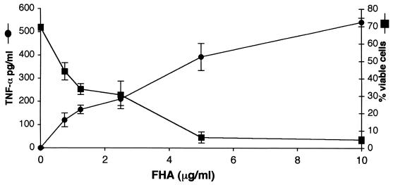
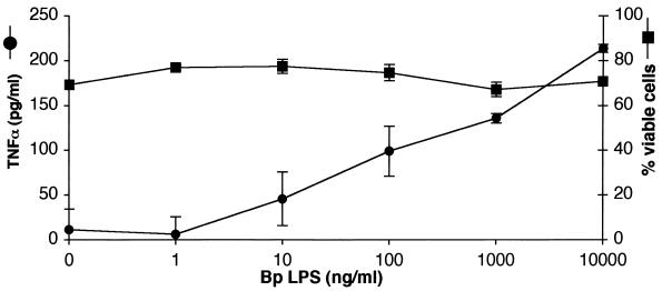
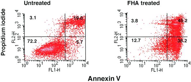
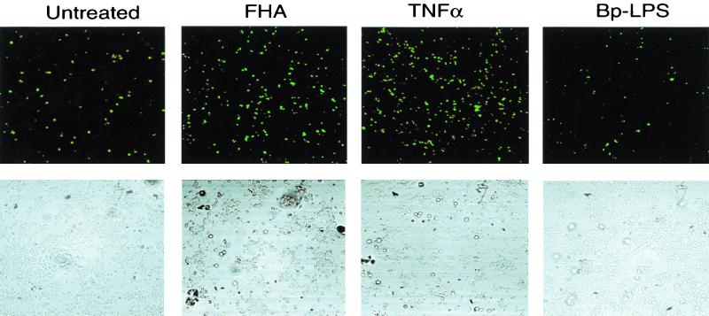
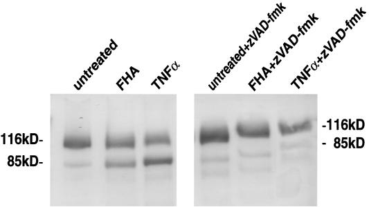
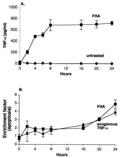
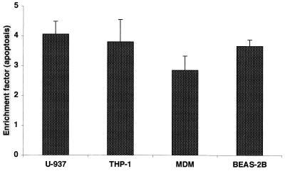
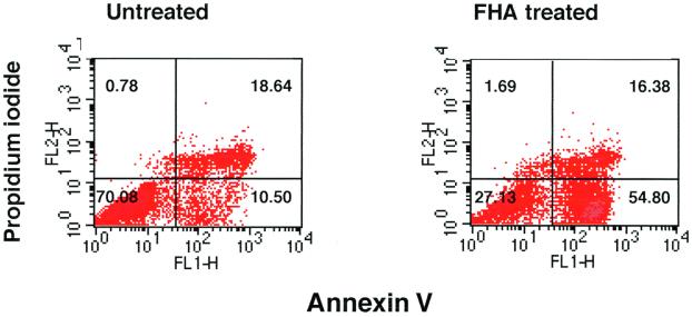
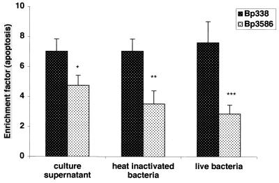
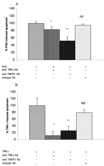
Filamentous hemagglutinin (FHA) is a dominant cell surface-associated Bordetella pertussis adhesin. Recognition that this protein is secreted in significant amounts and that bacterial adhesins may have other activities, prompted an assessment of FHA effects on human macrophages. Incubation of human macrophage-like U937 cells with preparations of FHA resulted in dose-dependent cytotoxicity, with death of 95% of treated cells after 24 h. Based on the use of four independent methods, death of these cells could be largely attributed to apoptosis. FHA-associated apoptosis was also observed in THP-1 macrophage-like cells, fresh human peripheral blood monocyte-derived macrophages (MDM), and BEAS-2B human bronchial epithelial cells. Infection of MDM with wild-type B. pertussis resulted in apoptosis within 6 h, while infection with an FHA-deficient derivative strain was only 50% as effective. FHA-associated cytotoxicity was preceded by host cell secretion of tumor necrosis factor alpha (TNF-alpha), a potential proapoptotic factor. However, pretreatment of cells with a neutralizing anti-TNF-alpha monoclonal antibody inhibited only 16% of the FHA-associated apoptosis. On the other hand, a blocking monoclonal antibody directed against TNF-alpha receptor 1 inhibited FHA-associated apoptosis by 47.7% (P = 0.0001), suggesting that this receptor may play a role in the death pathway activated by FHA. Our in vitro data indicate that secreted and cell-associated FHA elicits proinflammatory and proapoptotic responses in human monocyte-like cells, MDM, and bronchial epithelial cells and suggest a previously unrecognized role for this prominent virulence factor in the B. pertussis-host interaction.


Figures

FIG. 1
FIG. 1
FHA is associated with proinflammatory and cytotoxic responses in differentiated U937 cells. Cells were incubated for 20 h with various concentrations of FHA, and supernatants were then analyzed for secreted TNF-α (circles) and for the release of LDH (squares). The percentage of viable cells was calculated as described in Materials and Methods. Triplicate measurements were performed for each condition, and the standard error of the mean (bars) was calculated. These are representative data from one of four independent experiments with similar results.
FIG. 2
FIG. 2
B. pertussis LPS induces a proinflammatory response but not cell death. U937 cells were incubated for 20 h with various concentrations of B. pertussis (Bp) LPS. Supernatants were analyzed for TNF-α (circles) and LDH. The percentage of viable cells was calculated (squares). Triplicate measurements were performed for each condition, and the standard error of the mean (bars) was calculated. These are representative data from one of three independent experiments with similar results.
FIG. 3
FIG. 3
Annexin V-PI staining of U937 cells. Differentiated U937 cells were treated with medium only or with FHA (5 μg/ml) for 20 h and then stained with Annexin V-FITC–PI and analyzed by FACS (see Materials and Methods). These are representative data from one of three experiments with similar results.
FIG. 4
FIG. 4
Assessment of apoptosis in U937 cells using the TUNEL reaction. Cells were either untreated or incubated for 20 h with either FHA (5 μg/ml), TNF-α (50 ng/ml), or B. pertussis LPS (1 μg/ml). Labeled apoptotic nuclei were visualized with laser confocal microscopy at 488 nm (top row). The cells in the same field were also visualized with contrast microscopy (bottom row). These are representative fields from one of three experiments with similar results. Note that fewer cells remain intact after treatment with FHA and TNF-α than after other treatments (bottom row), due to programmed cell death.
FIG. 5
FIG. 5
Assessment of apoptosis by immunoblot analysis of PARP degradation. Differentiated U937 cells were pretreated for 1 h with either medium alone (left panel) or with zVAD-fmk (20 μg/ml) (right panel), and then incubated for 20 h in medium alone, FHA (5 μg/ml), or TNF-α (50 ng/ml). The cells were lysed and analyzed by immunoblotting with anti-PARP monoclonal antibody (see Materials and Methods). These are representative data from one of three experiments with similar results.
FIG. 6
FIG. 6
Time course of FHA-associated TNF-α secretion (A) and apoptosis (B) in U937 cells. Differentiated U937 cells were incubated for various periods of time with either medium alone (diamonds [panel A only]), FHA (5 μg/ml) (squares), or purified TNF-α (50 ng/ml) (circles [panel B only]) and then examined for TNF-α secretion (A) and for apoptosis (B) by ELISA methods (see Materials and Methods). The data shown are mean values from the analysis of samples in triplicate and are representative of three separate experiments. Bars indicate the standard error of the mean.
FIG. 7
FIG. 7
FHA-associated apoptosis in various human cell types. Differentiated U937 and THP-1 monocyte/macrophages, fresh peripheral blood MDMs, and BEAS-2B bronchial epithelial cells were incubated either with medium alone or FHA (5 μg/ml) for 20 h. Cells were then analyzed for apoptosis by an ELISA method. The data shown are mean values from the analysis of samples in triplicate and are representative of three separate experiments. Error bars indicate the standard error of the mean.
FIG. 8
FIG. 8
Annexin V-PI staining of fresh human MDMs. Cells were treated with medium only or with FHA (5 μg/ml) for 20 h and stained with Annexin V-FITC-PI and analyzed by FACS (see Materials and Methods). These are representative data from one of three experiments with similar results.
FIG. 9
FIG. 9
Apoptosis induced in macrophages by B. pertussis strains. Differentiated THP-1 monocytes were exposed for 20 h to culture supernatant from equal numbers of either B. pertussis BP 338 or BP 3586, or heat-inactivated bacteria at an MOI of 1:100. MDMs were exposed to live BP 338 or BP 3586 at an MOI of 1:10 for 6 h. Apoptosis was assessed by an ELISA method. The data shown are mean values from the analysis of samples in triplicate and are representative of three separate experiments. Error bars indicate the standard error of the mean. Statistical significance is indicated for comparisons between the two strains as analyzed by t test (two-sample test, assuming equal variances): P = 0.019 (∗), P = 0.04 (∗∗), and P = 0.01 (∗∗∗).
FIG. 10
FIG. 10
Inhibition of FHA-associated (A) and TNF-α-associated (B) apoptosis by anti-TNF-α and anti-TNFR1 blocking antibodies. Differentiated U937 cells were pretreated for 1 h with medium alone, anti-TNF-α antibody (10 μg/ml), anti-TNFR1 antibody (10 μg/ml), or an isotype-control antibody (10 μg/ml). Cells were then treated for 20 h with medium alone, FHA (5 μg/ml) (A), or TNF-α (50 ng/ml) (B). Cells were then analyzed for apoptosis by an ELISA method. The amount of apoptosis from cells without pretreatment was defined as 100%. The data shown are mean values from the analysis of samples in triplicate and are representative of three separate experiments. Bars indicate the standard error of the mean. Statistical significance was analyzed by t test as indicated for the comparisons between cells with and without pretreatment (two-sample test, assuming equal variances). (A) P = 0.044 (∗) and P = 0.0001 (∗∗); (B) P = 0.01 (∗) and P = 0.02 (∗∗). NS, not significant.

References

    1. Ashkenazi A, Dixit V M. Death receptors: signaling and modulation. Science. 1998;281:1305–1308. - PubMed
    1. Beutler B, Cerami A. Tumor necrosis, cachexia, shock, and inflammation: a common mediator. Annu Rev Biochem. 1988;57:505–518. - PubMed
    1. Boschwitz J S, Batanghari J W, Kedem H, Relman D A. Bordetella pertussis infection of human monocytes inhibits antigen-dependent CD4 T cell proliferation. J Infect Dis. 1997;176:678–686. - PubMed
    1. Cotter P A, Yuk M H, Mattoo S, Akerley B J, Boschwitz J, Relman D A, Miller J F. Filamentous hemagglutinin of Bordetella bronchiseptica is required for efficient establishment of tracheal colonization. Infect Immun. 1998;66:5921–5929. - PMC - PubMed
    1. Flak T A, Goldman W E. Signaling and cellular specificity of airway nitric oxide production in pertussis. Cell Microbiol. 1999;1:51–60. - PubMed

Publication types

Substances

LinkOut - more resources