

doi: 10.1128/IAI.69.4.2723-2727.2001.
Intracellular crystal formation as a mechanism of cytotoxicity in murine pulmonary Cryptococcus neoformans infection
Affiliations
- PMID: 11254641
- PMCID: PMC98213
- DOI: 10.1128/IAI.69.4.2723-2727.2001
Item in Clipboard
Intracellular crystal formation as a mechanism of cytotoxicity in murine pulmonary Cryptococcus neoformans infection
Infect Immun.
2001 Apr.
Abstract
Rod-like crystalline structures formed during eosinophilic Cryptococcus neoformans pneumonia in C57BL/6 mice. Crystals were found associated with yeast cells and free in host cell cytoplasm. The crystals apparently formed because of the interaction of a host protein with the cryptococcal polysaccharide. Crystal formation likely contributes to pathogenesis by causing cellular damage.
Figures
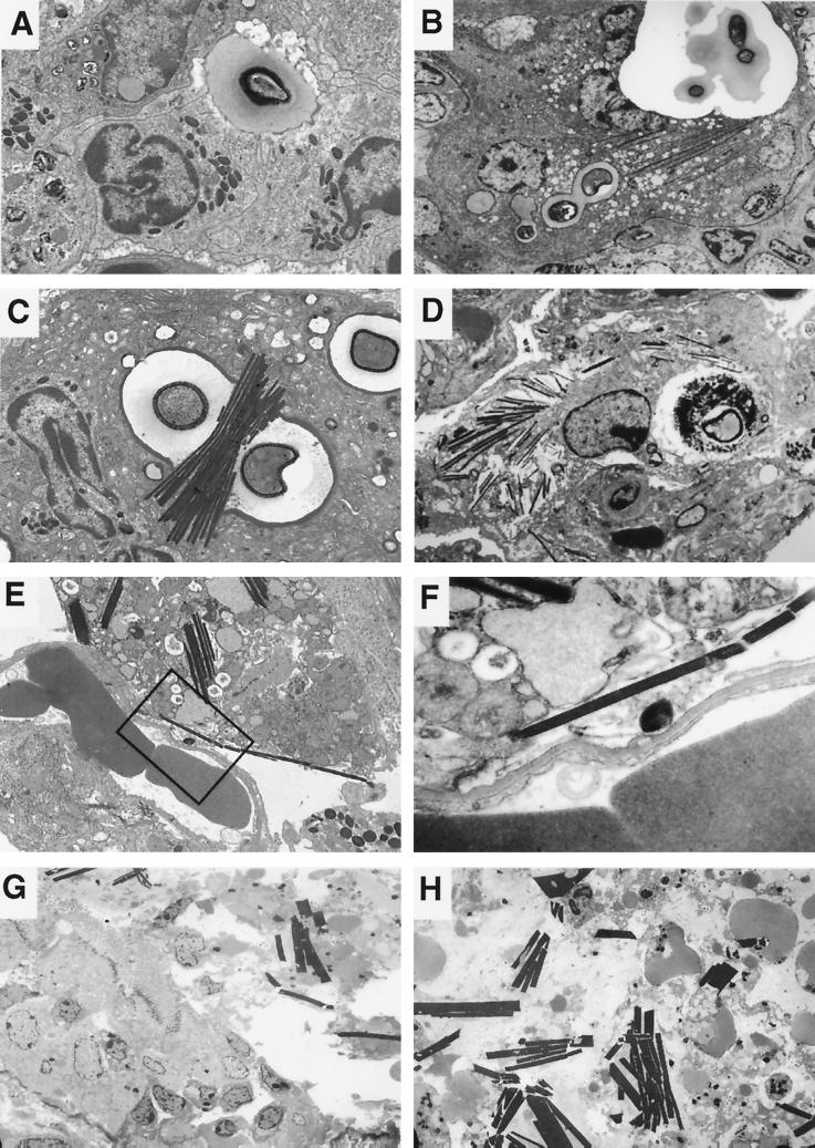
(A) Eosinophils recruited to the site of infection discharge electron-dense granular contents at the surface of an extracellular Cryptococcus, organism, allowing contact of cryptococcal polysaccharide with granule proteins (day 14; magnification, ×3,000). (B) The location of the yeast cell and crystals within a large multinucleated cell at a magnification lower than that for panel A. An electron-dense rim forms around the yeast cell, and crystals subsequently polymerize in contact with yeast and in the cytoplasm (day 14; magnification, ×2,000). (C) Budding intracellular C. neoformans in murine lung tissue 14 days after infection with electron-dense rim surrounding the polysaccharide capsule. Crystals formed in association with the yeast cell (magnification, ×4,000). (D) On day 28 after infection, large numbers of crystals were seen inside multinucleated cells, some of which were dying (magnification, ×2,000). (E) Crystals disrupted host cell membranes and became extracellular (day 28 after infection; magnification, ×3,000). (F) Higher magnification of the area enclosed in the boxed area in panel E demonstrates membrane disruption (magnification, ×20,000). (G and H) On day 28 after infection, crystals disrupted the bronchial epithelium, with loss of cilia. Epithelial cell debris and larger extracellular crystals were seen inside bronchi (magnification, ×1,000).
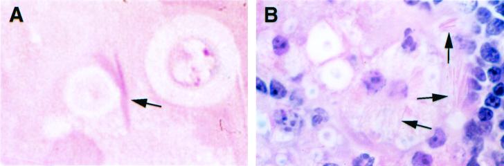
Light microscopic staining of intracellular crystals (arrows). (A) Crystals from a 1-μm-thick section of mouse lung 14 days after infection stained with eosin. (B) Staining of a 5-μm-thick section of mouse lung 28 days after infection with hematoxylin and eosin showing a macrophage containing a large number of intracellular crystals. Magnification, ×1,000 (panels A and B).
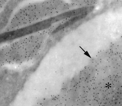
Immunoelctron microscopy shows the presence of gold label for cryptococcal polysaccharide surrounding the outer membrane of the crystals but not over the crystals. The asterisk is located on a cryptococcal capsule. The arrow points to artifactual contraction of the capsule from the edge of a phagosome. magnification, ×15,000).
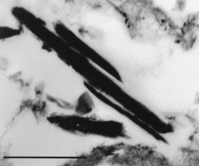
Crystals formed after incubation of C. neoformans strain 24067 with rat peritoneal inflammatory cells in vitro for 3 days. Bar, 1 μm.
Similar articles
-
IL-5 is required for eosinophil recruitment, crystal deposition, and mononuclear cell recruitment during a pulmonary Cryptococcus neoformans infection in genetically susceptible mice (C57BL/6).J Immunol. 1998 Mar 1;160(5):2393-400. J Immunol. 1998. PMID: 9498782
-
Role of granulocyte macrophage colony-stimulating factor in host defense against pulmonary Cryptococcus neoformans infection during murine allergic bronchopulmonary mycosis.Am J Pathol. 2007 Mar;170(3):1028-40. doi: 10.2353/ajpath.2007.060595. Am J Pathol. 2007. PMID: 17322386 Free PMC article.
-
Cryptococcus neoformans is a facultative intracellular pathogen in murine pulmonary infection.Infect Immun. 2000 Jul;68(7):4225-37. doi: 10.1128/IAI.68.7.4225-4237.2000. Infect Immun. 2000. PMID: 10858240 Free PMC article.
-
Pulmonary infection with capsule-deficient cryptococcus neoformans.Virchows Arch A Pathol Anat Histol. 1979 May 14;382(1):113-8. doi: 10.1007/BF01102745. Virchows Arch A Pathol Anat Histol. 1979. PMID: 157594
-
Proven pulmonary cryptococcosis due to capsule-deficient Cryptococcus neoformans does not differ clinically from proven pulmonary cryptococcosis due to capsule-intact Cr. neoformans.Mycoses. 2005 Jan;48(1):21-4. doi: 10.1111/j.1439-0507.2004.01068.x. Mycoses. 2005. PMID: 15679661
Cited by
-
Synergistic antifungal interaction of N-(butylcarbamothioyl) benzamide and amphotericin B against Cryptococcus neoformans.Front Microbiol. 2023 Mar 7;14:1040671. doi: 10.3389/fmicb.2023.1040671. eCollection 2023. Front Microbiol. 2023. PMID: 36960287 Free PMC article.
-
The influence of bacterial interaction on the virulence of Cryptococcus neoformans.Virulence. 2015;6(7):677-8. doi: 10.1080/21505594.2015.1088632. Virulence. 2015. PMID: 26364987 Free PMC article.
-
IL-4 dependent alternatively-activated macrophages have a distinctive in vivo gene expression phenotype.BMC Immunol. 2002 Jul 4;3:7. doi: 10.1186/1471-2172-3-7. BMC Immunol. 2002. PMID: 12098359 Free PMC article.
-
Role of host sphingosine kinase 1 in the lung response against Cryptococcosis.Infect Immun. 2010 May;78(5):2342-52. doi: 10.1128/IAI.01140-09. Epub 2010 Mar 1. Infect Immun. 2010. PMID: 20194596 Free PMC article.
-
Cryptococcal urease promotes the accumulation of immature dendritic cells and a non-protective T2 immune response within the lung.Am J Pathol. 2009 Mar;174(3):932-43. doi: 10.2353/ajpath.2009.080673. Epub 2009 Feb 13. Am J Pathol. 2009. PMID: 19218345 Free PMC article.
References
-
- Avrameas S. Coupling of enzymes to proteins with glutaraldehyde. Use of the conjugates for the detection of antigens and antibodies. Immunochemistry. 1969;6:43–52. - PubMed
-
- Casadevall A, Cleare W, Feldmesser M, Glatman-Freedman A, Goldman D L, Kozel T R, Lendvai N, Mukherjee J, Pirofski L-A, Rivera J, Rosas A L, Scharff M D, Valadon P, Westin K, Zhong Z. Characterization of a murine monoclonal antibody to Cryptococcus neoformans polysaccharide that is a candidate for human therapeutic studies. Antimicrob Agents Chemother. 1998;42:1437–1446. - PMC - PubMed
-
- Casadevall A, Mukherjee J, Devi S J N, Schneerson R, Robbins J B, Scharff M D. Antibodies elicited by a Cryptococcus neoformans-tetanus toxoid conjugate vaccine have the same specificity as those elicited in infection. J Infect Dis. 1992;165:1086–1093. - PubMed
Publication types
MeSH terms
Substances
Grants and funding
LinkOut - more resources
Full Text Sources
