. 2001 Mar;107(6):745-52.
doi: 10.1172/JCI11432.

Clonality and altered behavior of endothelial cells from hemangiomas

Affiliations

Clonality and altered behavior of endothelial cells from hemangiomas

E Boye et al. J Clin Invest. 2001 Mar.

Abstract

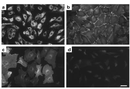
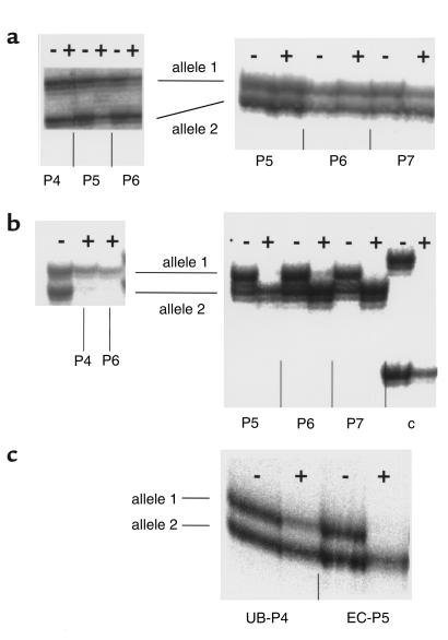
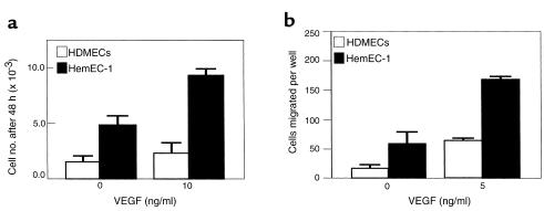
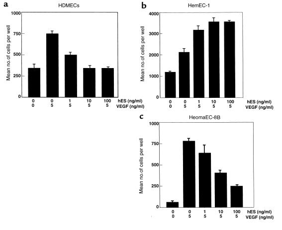
Hemangioma, the most common tumor of infancy, is a benign vascular neoplasm of unknown etiology. We show, for the first time to our knowledge, that endothelial cells from proliferating hemangioma are clonal, and we demonstrate that these hemangioma-derived cells differ from normal endothelial cells in their rates of proliferation and migration in vitro. Furthermore, migration of hemangioma endothelial cells is stimulated by the angiogenesis inhibitor endostatin, unlike the inhibition seen with normal endothelial cells. We conclude that hemangiomas constitute clonal expansions of endothelial cells. This is consistent with the possibility that these tumors are caused by somatic mutations in one or more genes regulating endothelial cell proliferation.


Figures

Figure 1
Figure 1
Characterization of hemangioma-derived endothelial cells by indirect immunofluorescence. Hemangioma-derived endothelial cells (hemEC-1) were fixed with methanol and incubated with anti-vWF (a), anti-CD31/PECAM-1 (b), anti–E-selectin (c), or isotype-matched control mouse IgG1 (d). Endothelial cells in c were activated with 0.2 ng/ml LPS for 5 hours to upregulate E-selectin (22). Bar, 10 μm.
Figure 2
Figure 2
Results of HUMARA assay on DNA from ECs of control infants’ skin, hemangioma lesions, and unbound fibroblast-like cells. Amplification was performed before (–) or after (+) HhaI digestion. For each cell sample shown, the individual is heterozygous for CAG repeat size and has two different alleles before HhaI digestion, represented by two major PCR products on gel. Fainter (“stutter”) bands are products of slippage by DNA polymerase in STR region. (a) Amplification of DNA from ECs cultured at P4 to P7 from skin of two control female infants HFSEC-3 (left) and HFSEC-11 (right). In each sample, two alleles are equally amplifiable both before and after digestion. (b) Amplification of DNA from cultured hemECs from patient 1 (left) at P4 and P6 and patient 21 (right) at P5, P6, and P7. In both cases, one allele completely disappears after HhaI digestion. This selective amplification of one allele indicates that it is always methylated and not subject to digestion by HhaI and, therefore, is the only inactive allele in cell population. C is a positive control for the HUMARA assay. (c) Amplification of DNA from cultured unbound, fibroblast-like cells of hemangioma 10 at P4 (UB-P4). For comparison, DNA from cultured hemEC-10 at P5 (EC-P5) is shown at right.
Figure 3
Figure 3
Comparison of in vitro properties of HDMECs vs. hemEC-1. (a) Comparison of proliferation rates of HDMECs vs. hemEC-1 in response to VEGF. HemEC-1 cells proliferate approximately 2.5-fold faster than HDMECs, both in the presence (10 ng/ml) or absence of exogenous VEGF. (b) Comparison of VEGF-induced migration of HDMECs vs. hemEC-1. HemEC-1 cells migrate approximately 3.5-fold faster than do HDMECs with (10 ng/ml) or without VEGF stimulation.
Figure 4
Figure 4
Effect of hES on VEGF-induced migration of HDMECs, hemEC-1, and heomaEC-8B. (a) Migration of HDMECs is inhibited by hES. Complete inhibition is achieved at approximately 10 ng/ml hES. (b) HemEC-1 cells exhibit the opposite response to the other cells; their migration is strongly stimulated by hES even at 1 ng/ml. (c) Migration of heomaEC-8B is inhibited by hES.

Comment in

  • Pathogenesis of hemangioma.
    Marchuk DA. Marchuk DA. J Clin Invest. 2001 Mar;107(6):665-6. doi: 10.1172/JCI12470. J Clin Invest. 2001. PMID: 11254664 Free PMC article. Review. No abstract available.

References

    1. Mulliken, J.B. 1988. Diagnosis and natural history of hemangiomas. In Vascular birthmarks: hemangiomas and malformations. J.B. Mulliken and A.E. Young, editors. W.B. Saunders Co. Philadelphia, Pennsylvania, USA. 41–62.
    1. Razon MJ, Kräling BM, Mulliken JB, Bischoff J. Increased apoptosis coincides with onset of involution in infantile hemangioma. Microcirculation. 1998;5:189–195. - PubMed
    1. Mulliken, J.B., Fishman, S.J., and Burrows, P.E. 2000. Vascular anomalies. In Current problems in surgery. S.A. Wells and L.L. Creswell, editors. Mosby. St. Louis, Missouri, USA. 517–584. - PubMed
    1. Berard M, et al. Vascular endothelial growth factor confers a growth advantage in vitro and in vivo to stromal cells cultured from neonatal hemangiomas. Am J Pathol. 1997;150:1315–1326. - PMC - PubMed
    1. Kräling BM, Bischoff J. A simplified method for growth of human microvascular endothelial cells results in decreased senescence and continued responsiveness to cytokines and growth factors. In Vitro Cell Dev Biol Anim. 1998;34:308–315. - PubMed

Publication types

MeSH terms