. 2001 Mar;107(6):R37-45.
doi: 10.1172/JCI11297.

CCR6-deficient mice have impaired leukocyte homeostasis and altered contact hypersensitivity and delayed-type hypersensitivity responses

Affiliations

CCR6-deficient mice have impaired leukocyte homeostasis and altered contact hypersensitivity and delayed-type hypersensitivity responses

R Varona et al. J Clin Invest. 2001 Mar.

Abstract

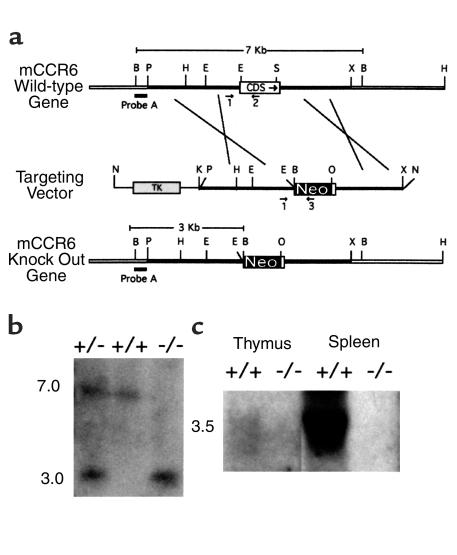
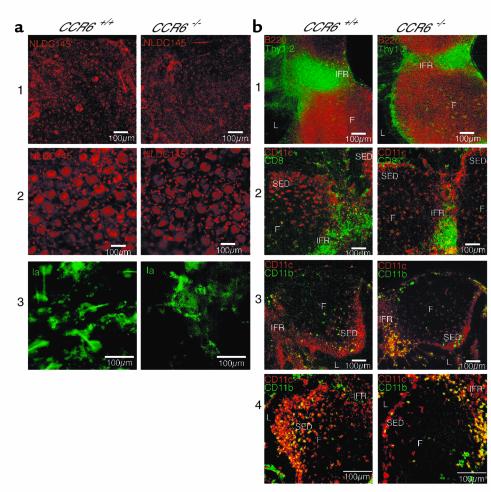
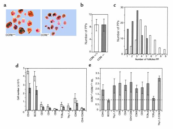
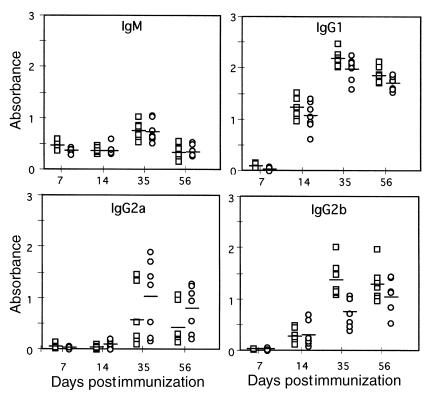
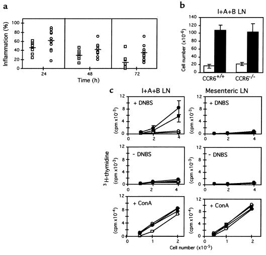
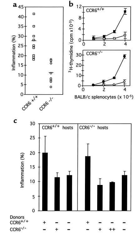
CCR6 expression in dendritic, T, and B cells suggests that this beta-chemokine receptor may regulate the migration and recruitment of antigen-presenting and immunocompetent cells during inflammatory and immunological responses. Here we demonstrate that CCR6-/- mice have underdeveloped Peyer's patches, in which the myeloid CD11b+ CD11c+ dendritic-cell subset is not present in the subepithelial dome. CCR6-/- mice also have increased numbers in T-cell subpopulations within the intestinal mucosa. In 2,4-dinitrofluorobenzene-induced contact hypersensitivity (CHS) studies, CCR6-/- mice developed more severe and more persistent inflammation than wild-type (WT) animals. Conversely, in a delayed-type hypersensitivity (DTH) model induced with allogeneic splenocytes, CCR6-/- mice developed no inflammatory response. The altered responses seen in the CHS and DTH assays suggest the existence of a defect in the activation and/or migration of the CD4(+) T-cell subsets that downregulate or elicit the inflammation response, respectively. These findings underscore the role of CCR6 in cutaneous and intestinal immunity and the utility of CCR6-/- mice as a model to study pathologies in these tissues. This article was published online in advance of the print edition. The date of publication is available from the JCI website, http://www.jci.org.


Figures

Figure 1
Figure 1
Targeted disruption of the CCR6 gene. (a) Targeting strategy. CCR6 WT locus with partial restriction map. The coding sequence (CDS), neomycin resistance gene (Neo), and thymidine kinase gene (TK) are shown as open, filled, and shaded boxes, respectively. A thick filled bar shows probe A, used in Southern blot analysis for screening genomic DNA. Arrows mark the position and direction of synthesis of oligonucleotides used in PCR genotyping. B, BamHI; P, PstI; H, HindIII; E, EcoRI; S, SmaI; X, XbaI; N, NotI; K, KpnI; O, XhoI. (b) Representative Southern blot analysis of BamHI-digested tail DNA from WT (+/+), heterozygous (+/–), and homozygous (–/–) CCR6 mice using probe A. Band sizes in kilobases for the WT and knockout alleles are indicated on the left. (c) Representative Northern blot analysis of the CCR6 mRNA expression in thymus and spleen from CCR6 WT (+/+) and knockout (–/–) animals. The size of the CCR6 transcript in kilobases is indicated on the left.
Figure 2
Figure 2
Similar LC complements in the skin, but altered positioning of the myeloid DC subpopulation within the PPs of CCR6–/– mice. (a) Samples of skin epidermis (panel 1) and dermis (panel 2) were stained with the NLDC145 Ab and analyzed by fluorescence microscopy. In panel 3, dorsal ear skin preparations were cultured for 72 hours in RPMI-1640. Dermal sheets were then separated from epidermis and stained with anti-Iab to reveal the existence of LC cords. (b) Staining of WT (CCR6+/+) and knockout (CCR6–/–) mouse PPs with anti-B220 (red) and anti-Thy1.2 (green) reveals B cells in follicles (F) and T cells in the IFR. For orientation, lumen (L) position is indicated. In panel 2, staining with anti-CD11c (red) and anti-CD8α (green) localizes lymphoid DC mainly in the IFR. In panels 3 and 4, staining with anti-CD11c (red) and anti-CD11b (green) shows a defect in the positioning of myeloid DC in CCR6–/– mice, where they are located mainly in the IFR and not in SED. Scale bars = 100 μm.
Figure 3
Figure 3
CCR6–/– mice have underdeveloped PPs and impaired lymphocyte homeostasis in the intestinal mucosa. (a) Low-magnification micrography of dissected PPs from 4-month-old WT (CCR6+/+) and CCR6–/– mice showing the different level of development of these lymphoid organs. (b) The number of PPs is similar in WT (CCR6+/+) and CCR6–/– mice. Data shown correspond to the average number found in ten animals of each genotype. (c) The number of developed follicles per patch and the number of PPs with a given developmental state differ in CCR6–/– mice (filled bars) from those of WT animals (open bars). Accumulated data are presented from ten animals per group. (d) Flow-cytometry analysis of lymphocyte subsets in PPs of WT (open bars) and CCR6–/– mice (filled bars). (e) The cell numbers in IEL subpopulations are increased in CCR6–/– mice (n = 2–3 pooled animals of each genotype in each experiment). Data shown in d and e correspond to the mean and SE from five independent experiments.
Figure 4
Figure 4
Serum concentrations of antigen-specific immunoglobulins in animals immunized with DNP-KLH. CCR6–/– (n = 7; circles) and CCR6+/+ (n = 7; squares) mice were immunized subcutaneously with 30 μg of DNP-KLH in CFA and boosted twice with 30 μg of DNP-KLH in IFA at days 10 and 28 postimmunization. Mice were bled from the retro-orbital plexus at days 7 and 14, then every 21 days, and serum concentration of DNP-KLH–specific Ig isotypes were determined using ELISA. Individual data from the 10–3 dilution and the mean value for each group are presented. Two-tailed t-test value for the IgG2b data at day 35 is P = 0.0032; P > 0.05 for the rest.
Figure 5
Figure 5
Altered response of CCR6–/– mice in contact hypersensitivity inflammation. (a) CCR6–/– (n = 12; circles) and CCR6+/+ (n = 12; squares) mice were sensitized by epicutaneous application of 25 μl of 0.5% DNFB solution on the shaved abdomen. Increases in ear swelling were measured 24, 48, and 72 hours after challenge with 20 μl of 0.2% DNFB on the ears. Individual data and the mean value for each group are presented. Two-tailed t-test values for DNFB data: P = 0.006 (24 hours), P = 0.005 (48 hours), P = 0.002 (72 hours). (b) Similar number of lymph node cells in CCR6+/+ and CCR6–/– mice. Animals were sensitized with DNFB (filled bars) or left untreated (open bars); 5 days later, lymphocytes from IAB (I+A+B) LNs were prepared and counted. (c) CCR6–/– lymphocytes proliferate in response to specific and nonspecific stimuli. CCR6–/– and CCR6+/+ animals were sensitized with DNFB (filled symbols) or untreated (open symbols); 5 days later, cells from draining (I+A+B) or control mesenteric LNs were recovered and cultured for 36 hours alone or in the presence of DNBS, as indicated. Cultures were pulsed with 3H-thymidine for 18 hours, and cell proliferation was estimated. Similar experiments were performed to measure in vitro responses to a polyclonal stimulus, Con A. Each value represents the average of three separate groups, consisting of pooled LNs from two mice. All assays were performed in triplicate.
Figure 6
Figure 6
CCR6–/– mice have a markedly diminished DTH response to allogeneic BALB/c splenocytes. (a) CCR6–/– (n = 10; circles) and CCR6+/+ (n = 11; squares) animals were sensitized by intravenous injection of 106 BALB/c splenocytes. Five days later, mice were challenged by injecting 13 × 106 BALB/c splenocytes into their right footpads, and swelling was measured 24 hours later. Individual data and the mean value for each group are presented. The two-tailed t-test value for these data was P = 0.000016. (b) In vitro MLR to allogeneic BALB/c splenocytes. Lymphocytes were prepared from IAB LNs from CCR6+/+ (filled squares) and CCR6–/– mice (filled circles) and cultured in 96-well plates (2 × 10⁵ cells/well) with increasing amounts of stimulator allogeneic BALB/c splenocytes. After 72 hours, cultures were pulsed with 3H-thymidine for 24 hours and cell proliferation was estimated. Background proliferation is also shown (open symbols). Each point represents the average value of two separate groups, each consisting of pooled LNs from two mice. Assays were performed in triplicate. (c) Adoptive transfer of sensitized CCR6+/+ CD4+ T cells to CCR6–/– mice restores their ability to produce a DTH response. Unsensitized CCR6+/+ and CCR6–/– mice were adoptively transferred with 2 × 10⁷ CD4+ T cell–enriched preparations purified from sensitized CCR6+/+ and CCR6–/– donors, as indicated. T cells from CCR6–/– IAB LNs were also allowed to proliferate in an in vitro MLR assay with BALB/c splenocytes before being injected to CCR6–/– hosts (++). After 16 hours, animals were challenged with 13 × 106 BALB/c splenocytes in footpads, and swelling was measured 24 hours later.

References

    1. Banchereau J, et al. Immunobiology of dendritic cells. Annu Rev Immunol. 2000;18:767–811. - PubMed
    1. Forster R, et al. A putative chemokine receptor, BLR1, directs B cell migration to defined lymphoid organs and specific anatomic compartments of the spleen. Cell. 1996;87:1037–1047. - PubMed
    1. Sallusto F, Lanzavecchia A, Mackay CR. Chemokines and chemokine receptors in T-cell priming and Th1/Th2-mediated responses. Immunol Today. 1998;19:568–574. - PubMed
    1. Sallusto F, Lenig D, Forster R, Lipp M, Lanzavecchia A. Two subsets of memory T lymphocytes with distinct homing potentials and effector functions. Nature. 1999;401:708–712. - PubMed
    1. Forster R, et al. CCR7 coordinates the primary immune response by establishing functional microenvironments in secondary lymphoid organs. Cell. 1999;99:23–33. - PubMed

Publication types

MeSH terms