. 2001 May;55(2):242-53.
doi: 10.1002/1097-4636(200105)55:2<242::aid-jbm1011>3.0.co;2-d.

Bone tissue engineering in a rotating bioreactor using a microcarrier matrix system

Affiliations

Bone tissue engineering in a rotating bioreactor using a microcarrier matrix system

E A Botchwey et al. J Biomed Mater Res. 2001 May.

Abstract

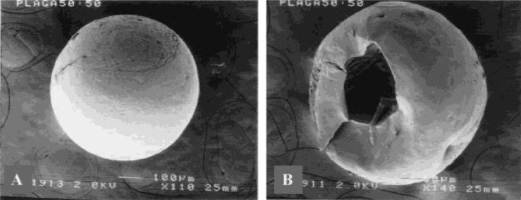
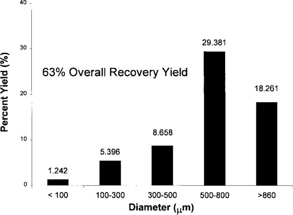
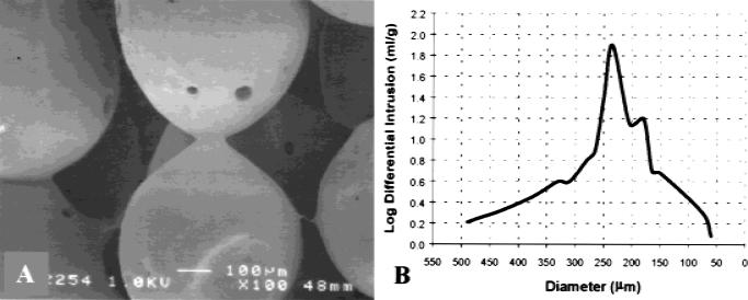
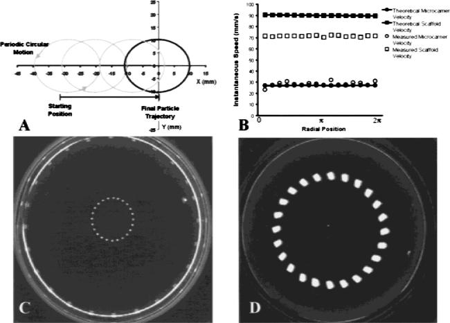
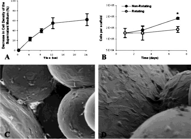
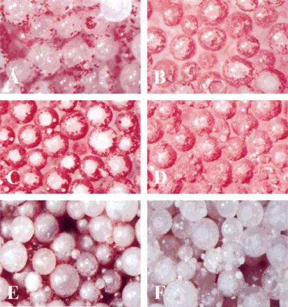
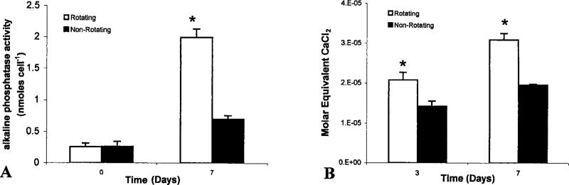
A novel approach was utilized to grow in vitro mineralized bone tissue using lighter-than-water, polymeric scaffolds in a high aspect ratio rotating bioreactor. We have adapted polymer microencapsulation methods for the formation of hollow, lighter-than-water microcarriers of degradable poly(lactic-co-glycolic acid). Scaffolds were fabricated by sintering together lighter-than-water microcarriers from 500 to 860 microm in diameter to create a fully interconnected, three-dimensional network with an average pore size of 187 microm and aggregate density of 0.65 g/mL. Motion in the rotating bioreactor was characterized by numerical simulation and by direct measurement using an in situ particle tracking system. Scaffold constructs established a near circular trajectory in the fluid medium with a terminal velocity of 98 mm/s while avoiding collision with the bioreactor wall. Preliminary cell culture studies on these scaffolds show that osteoblast-like cells readily attached to microcarrier scaffolds using controlled seeding conditions with an average cell density of 6.5 x 10(4) cells/cm(2). The maximum shear stress imparted to attached cells was estimated to be 3.9 dynes/cm(2). In addition, cells cultured in vitro on these lighter-than-water scaffolds retained their osteoblastic phenotype and showed significant increases in alkaline phosphatase expression and alizarin red staining by day 7 as compared with statically cultured controls.


Figures

Figure 1
Figure 1
SEM micrograph of hollow microcarrier morphology. (A) An intact hollow microcapsule of PLAGA 50:50 (original magnification x110). (B) Hollow internal morphology is confirmed by visualization after immersion and fracture in liquid N2 (original magnification x140).
Figure 2
Figure 2
Size distribution and percent yield of hollow microcarriers. Yield is defined as the fraction of starting raw material (PLAGA) that is recovered in the form of lighter than-water microcarriers after fabrication.
Figure 3
Figure 3
Characterization of lighter-than-water scaffolds by SEM and mercury porosimetry. (A) SEM micrograph of polymer scaffold formed by the sintered microsphere method, using spheres with diameters between 500–860 μm. The sintered microspheres created an interconnected, porous structure suitable for cellular proliferation and tissue ingrowth (original magnification x100). (B) A plot log differential mercury intrusion versus pore diameter illustrates typical pore size distribution of lighter-than-water composed of 500–860-μm hollow microcarriers.
Figure 4
Figure 4
Lighter-than-water microcarrier and scaffold motion in the HARV rotating bioreactor. (A) Theoretical prediction of the trajectory for a 600-μm hollow PLAGA microcarrier in the rotating bioreactor at 25 rpm (density 0.65 g/mL). (B) Comparison of a single microcarrier and a single scaffold instantaneous speed. Single microcarrier data for a 600-μm particle with density 0.65 g/mL is in good agreement with theory. Microcarrier scaffold data for a 4 × 2.5 mm, 0.65 g/mL scaffold deviates significantly from the theoretical prediction for a uniform sphere of similar size and density. (C) Trajectory of a 600-μm hollow PLAGA microcarrier in the rotating bioreactor turning at 25 rpm. Consecutive frames were recorded and digitized to render a temporal description microcarrier motion. The diameter of the surrounding vessel wall is approximately 100 mm. (D) Trajectory of a 4 × 2.5 mm scaffold in the rotating bioreactor turning at 25 rpm. The scaffold is composed of 500–860-μm hollow PLAGA microcarriers and has an aggregate density of 0.65 g/mL.
Figure 5
Figure 5
Analysis of cell attachment and growth during bioreactor rotation. (A) Cell concentration decreased by 60% on average after 24 h of co-inoculation with lighter-than-water scaffolds in the rotating bioreactor, and initial cell attachment was calculated accordingly. (B) DNA cell counting to confirm initial cell seeding was in good agreement with estimates determined from cell concentration. Significantly, fewer cells were detected on lighter-than-water scaffolds cultured in a rotating bioreactor (25 rpm) by 7 days were by day as compared with nonrotated 3-D controls (asterisk denotes significance obtained at 5% type I error and n = 3). (C,D) SEM micrographs (original magnification x150 and x300, respectively) of sintered microsphere scaffold cultured with SaOS osteoblasts cells for 7 days. The cells were found on and between the microspheres. The cells bridged the microspheres and had completely covered the spheres. Cell extensions were found throughout the structure.
Figure 6
Figure 6
Light micrographs (original magnification x50) showing phenotypic expression and early mineralized matrix synthesis by osteoblast-like cells in the rotating bioreactor. (A–D) Alkaline phosphatase expression by osteoblast-like cells cultured under rotating conditions at days 3 (A) and 7 (C). Right panels show companion micrographs of alkaline phosphatase expression on nonrotated 3-D controls at days 3 (B) and 7 (D). (E–F) Lighter-than-water scaffolds were cultured in the bioreactor for 7 days under static conditions (E) and 25 rpm rotation (F). Samples were stained with ALZ to detect mineralized extracellular matrix. Substantially more alizarin-positive material was observable on 3-D scaffolds cultured under rotating conditions.
Figure 7
Figure 7
Colorimetric determination of alkaline phosphatase expression and mineralized matrix synthesis. (A) Aliquots of individual scaffold homogenate were diluted to a known concentration and analyzed for alkaline phosphatase expression. Optical density measurements were determined at 415 nm. Quantities of alkaline phosphatase were normalized by dividing the measured quantity by the total number of cells present on the scaffold from which the homogenate was prepared. (B) To quantify the amount of ALZ that reacts with mineralized matrix, we adapted a technique that solubilizes the red matrix precipitate with cetyl pyridinium chloride to yield a purple solution suitable for optical density measurements at 562 nm. The concentration of Ca2+ equivalents in scaffold homogenate was determined by using a CaCl2 standard. For each time point, molar equivalent quantities were normalized for the average number of cells per scaffold as determined by companion fluorometric DNA analysis.

References

    1. Langer R, Vacanti JP. Tissue engineering. Science. 1993;260(5110):920–926. - PubMed
    1. Langer R, Vacanti JP, Vacanti C, Atala A, Freed LE, Vunjak-Novakovic G. Tissue engineering: Biomedical applications. Tissue Eng. 1995;1(2):151–161. - PubMed
    1. Burwell RG. History of bone grafting and bone substitutes with special reference to osteogenic induction. In: Urist MR, Burwell RG, editors. Bone grafts, derivatives, and substitutes. Butterworth-Heinemann; Oxford: 1994. p. 3.
    1. Cook SD, Baffes GC, Wolfe MW, Sampath TK, Rueger DC, Whitecloud TS. The effect of recombinant human osteogenic protein-1 on healing of large segmental bone defects. J Bone Joint Surg Am. 1994;76(6):827. - PubMed
    1. Gazdag AR, Lane JM, Glaser D, Forster RA. Alternative to autogenous bone graft: Efficacy and indications. J Am Acad OrthoP Surg. 1995;3(1):1. - PubMed

Publication types

LinkOut - more resources