

Review
doi: 10.1093/embo-reports/kvd001.
The G x U wobble base pair. A fundamental building block of RNA structure crucial to RNA function in diverse biological systems
Affiliations
- PMID: 11256617
- PMCID: PMC1083677
- DOI: 10.1093/embo-reports/kvd001
Item in Clipboard
Review
The G x U wobble base pair. A fundamental building block of RNA structure crucial to RNA function in diverse biological systems
EMBO Rep.
2000 Jul.
Abstract
The G x U wobble base pair is a fundamental unit of RNA secondary structure that is present in nearly every class of RNA from organisms of all three phylogenetic domains. It has comparable thermodynamic stability to Watson-Crick base pairs and is nearly isomorphic to them. Therefore, it often substitutes for G x C or A x U base pairs. The G x U wobble base pair also has unique chemical, structural, dynamic and ligand-binding properties, which can only be partially mimicked by Watson-Crick base pairs or other mispairs. These features mark sites containing G x U pairs for recognition by proteins and other RNAs and allow the wobble pair to play essential functional roles in a remarkably wide range of biological processes.
Figures
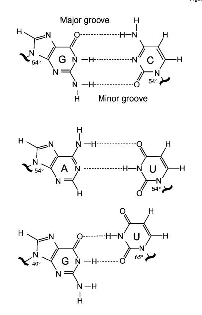
Fig. 1. Watson–Crick G·C and A·U base pairs differ from wobble G·U pairs in the type and location of functional groups that are projected into major and minor grooves. These pairs also differ in the orientation of the bases with respect to the phosphodiester backbone. Whereas the glycosidic angle is similar (~54°) for all nucleosides in Watson–Crick pairs, both angles for G and U differ in the wobble pair.
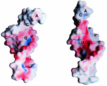
Fig. 2. The G·C, A·U and G·U pairs have different functional groups in the major groove, leading to a significantly more electronegative environment at and near the G·U pair. As shown in this image, the surface electrostatic potential (negative is red and positive is blue) of tRNAAla acceptor end minihelices differs between the wild-type molecule containing G·U (right) and a mutant molecule where G·U is replaced by G·C (left).
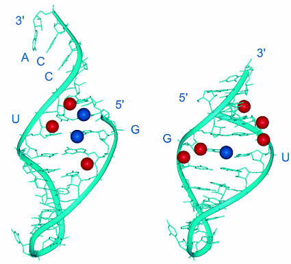
Fig. 3. View into the minor groove of RNA double helices showing the exocyclic amino groups (blue) and 2′-OH groups (red) that are critical for the activity of the G·U pairs in different functional contexts. The figure shows the three-dimensional structure of the tRNAAla acceptor end minihelix (left) and of the P1 helix substrate of group I self-splicing introns (right), highlighting chemical groups in the minor groove that contribute to G·U activity. A set of related but not identical chemical groups at and around the wobble pair is essential for activity in both contexts.
References
-
- Abramovitz D.L., Friedman, R.A. and Pyle, A.M. (1996) Catalytic role of 2′-hydroxyl groups within a group II intron active site. Science, 271, 1410–1413. - PubMed
-
- Allain F.H.-T. and Varani, G. (1995b) Structure of the P1 helix from group I self splicing introns. J. Mol. Biol., 250, 333–353. - PubMed
-
- Cate J.H. and Doudna, J.A. (1996) Metal-binding sites in the major groove of a large ribozyme domain. Structure, 4, 1221–1229. - PubMed
Publication types
MeSH terms
Substances
LinkOut - more resources
Full Text Sources
Other Literature Sources
