. 2001 Mar 19;152(6):1123-34.
doi: 10.1083/jcb.152.6.1123.

UGO1 encodes an outer membrane protein required for mitochondrial fusion

Affiliations

UGO1 encodes an outer membrane protein required for mitochondrial fusion

H Sesaki et al. J Cell Biol. .

Erratum in

  • J Cell Biol 2001 Apr 30;153(3):635

Abstract

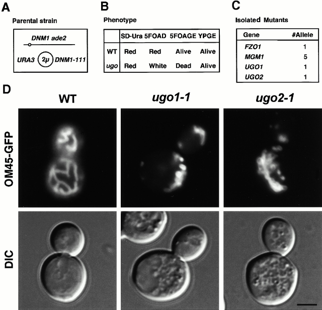
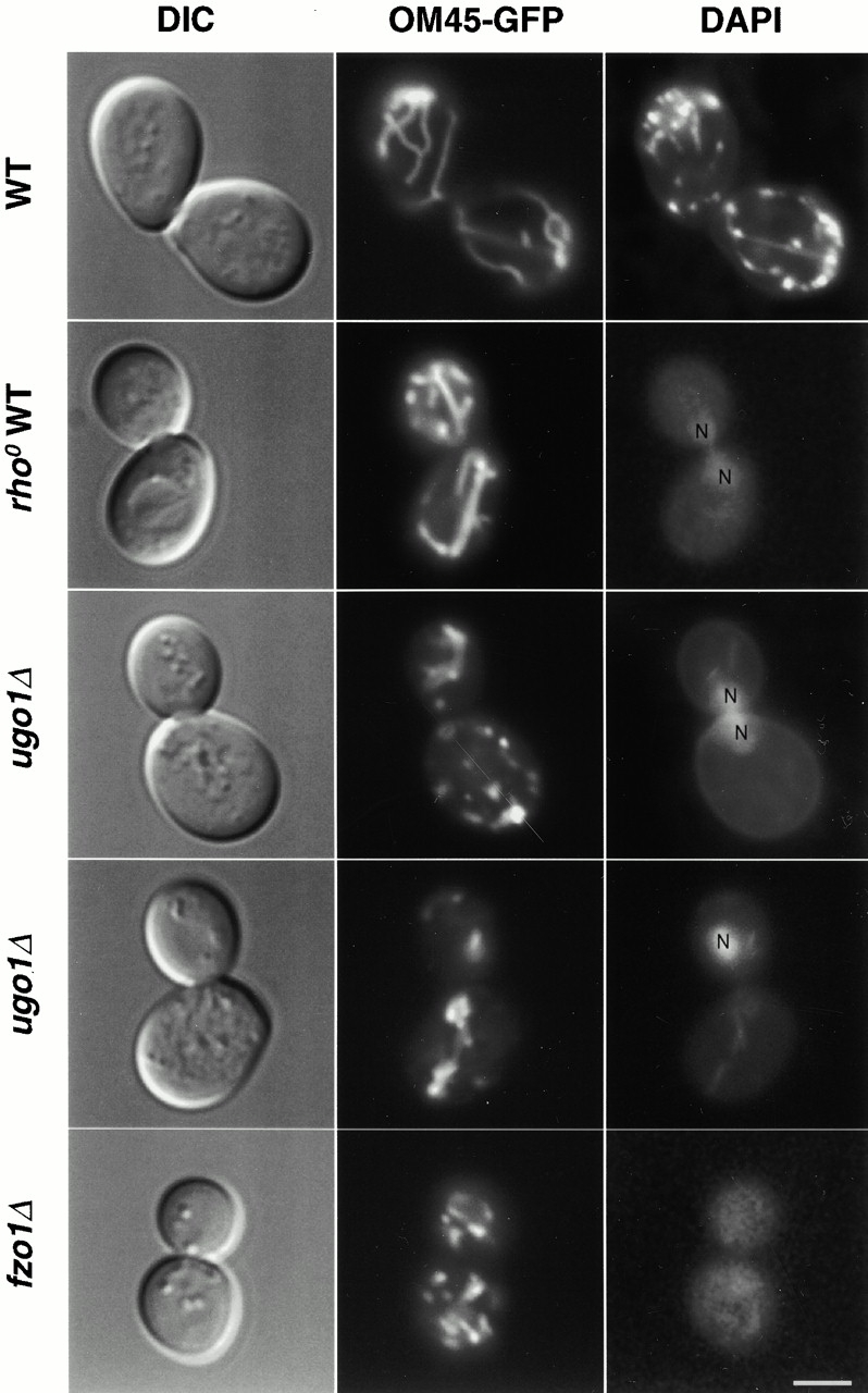
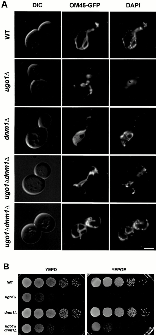
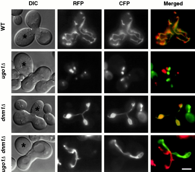
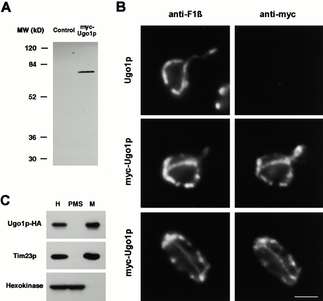
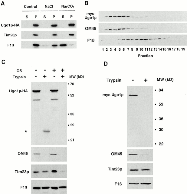
Membrane fusion plays an important role in controlling the shape, number, and distribution of mitochondria. In the yeast Saccharomyces cerevisiae, the outer membrane protein Fzo1p has been shown to mediate mitochondrial fusion. Using a novel genetic screen, we have isolated new mutants defective in the fusion of their mitochondria. One of these mutants, ugo1, shows several similarities to fzo1 mutants. ugo1 cells contain numerous mitochondrial fragments instead of the few long, tubular organelles seen in wild-type cells. ugo1 mutants lose mitochondrial DNA (mtDNA). In zygotes formed by mating two ugo1 cells, mitochondria do not fuse and mix their matrix contents. Fragmentation of mitochondria and loss of mtDNA in ugo1 mutants are rescued by disrupting DNM1, a gene required for mitochondrial division. We find that UGO1 encodes a 58-kD protein located in the mitochondrial outer membrane. Ugo1p appears to contain a single transmembrane segment, with its NH(2) terminus facing the cytosol and its COOH terminus in the intermembrane space. Our results suggest that Ugo1p is a new outer membrane component of the mitochondrial fusion machinery.


Figures

Figure 1
Figure 1
Isolation of ugo mutants. (A) Strain used to isolate ugo1 and ugo2. ade2 DNM1 strains that carry plasmid pHS50, which expresses the dominant negative Dnm1-111 protein, lack Dnm1p activity. Cells that lose pHS50 contain functional Dnm1p. (B) Mutant isolation scheme. The parental strain (WT) maintains mtDNA on glucose-containing medium in the presence (SD-Ura) or absence (5FOAD) of the URA3-DNM1-111 plasmid and form red colonies due to the ade2 mutation (Reaume and Tatum 1949). ugo mutants maintain mtDNA only in the presence of the pHS50, forming red colonies on SD-Ura, but become white on 5FOAD medium, which selects for loss of the URA3-DNM1-111 plasmid (Boeke et al. 1984). Growth on glycerol and ethanol medium confirms that ugo mutants contain mtDNA in the presence of the URA3-DNM1-111 plasmid (YPGE), but that ugo mutants are inviable on 5FOA medium containing glycerol and ethanol (5FOAGE). (C) Four different genes were identified in the ugo screen. Complementation tests showed one fzo1 mutant, five mgm1 mutants, and two new mutants, ugo1 and ugo2, were isolated. (D) ugo mutants contain highly fragmented mitochondria. Wild-type (W303), ugo1-1 (YHS64), and ugo2-1 (YHS65) were grown on 5FOAD medium to select for cells that lost the URA3-DNM1-111 plasmid, pHS50, and then transformed with pKC2, which expresses GFP fused to the COOH terminus of the mitochondrial outer membrane protein, OM45p (OM45-GFP; Cerveny et al. 2001). Cells were grown to log phase in SRaf medium and examined by fluorescence microscopy. Fluorescence (OM45-GFP) and DIC images are shown. Bar, 3 μm.
Figure 2
Figure 2
ugo1Δ cells grow slowly on glucose-containing medium and are inviable on nonfermentable carbon sources. (A) 10 meiotic products from the ugo1::HIS3/UGO1 diploid strain, YHS91, were separated by micromanipulation and allowed to grow at 30°C for 5 d on YEPD. (B) 10 tetrads from the sporulated ugo1::HIS3/UGO1 diploid strain, YHS91, were patched onto YEPD medium and then replica-plated to SD medium lacking histidine (SD-His) and YEPGE. Cells were incubated at 30°C for 6 d.
Figure 3
Figure 3
ugo1Δ cells contain fragmented mitochondria and lack mtDNA. Wild-type (FY833), rho0 WT (YHS92), and ugo1Δ (YHS72) and fzo1Δ (YHS74) cells were transformed with OM45-GFP–expressing plasmid, pKC2 (Cerveny et al. 2001). Cells were grown to log phase in SRaf medium. Cells were stained using 1 μg/ml DAPI, and viewed by DIC and fluorescence (OM45-GFP or DAPI) microscopy. rho0 cells (YHS92) were generated by treating wild-type cells (FY833) with 25 μg/ml ethidium bromide as described (Fox et al. 1991). N, nuclear DNA staining. Bar, 3 μm.
Figure 4
Figure 4
Morphology and distribution of vacuoles, the endoplasmic reticulum, and the actin cytoskeleton are normal in ugo1Δ cells. (A) Wild-type (FY833, WT) and ugo1Δ (YHS72) cells were grown to log phase in YEPD medium. Cells were then stained with 12 μM FM4-64 (Molecular Probes) to label vacuoles (Vida and Emr 1995) and examined by fluorescence (FM4-64) and DIC microscopy. (B) Wild-type and ugo1Δ cells expressing GFP fused to the COOH terminus of Sec63p from pPS1530 (Prinz et al. 2000) were grown to log phase in SD medium and examined by fluorescence microscopy. (C) Wild-type and ugo1Δ cells were grown to log phase in YEPGal medium. Cells were then fixed in 3.7% formaldehyde and stained with 1 μM Alexa 594–phalloidin (Molecular Probes) to visualize actin filaments (Adams and Pringle 1991). Bars, 3 μm.
Figure 5
Figure 5
ugo1Δ dnm1Δ cells contain mitochondrial tubules and maintain mtDNA. Wild-type (FY833, WT), ugo1Δ (YHS72), dnm1Δ (YHS83), and ugo1Δ dnm1Δ (YHS85) cells expressing OM45-GFP (pKC2) were grown to log phase in SRaf medium, stained with 1 μg/ml DAPI, and viewed by DIC and fluorescence (OM45-GFP) microscopy. (B) Wild-type, ugo1Δ, dnm1Δ, and ugo1Δ dnm1Δ cells were grown to log phase in YEPD medium. Cells were collected and resuspended in YEPD medium to an OD600 of 2. Cells were then diluted in 10-fold increments, and 10 μl of each dilution was spotted onto YEPD and YEPGE media and incubated at 30°C for 2 and 6 d, respectively. N, nuclear DNA staining. Bar, 3 μm.
Figure 6
Figure 6
Mitochondrial fusion is defective in ugo1Δ and ugo1Δ dnm1Δ cells. MATa cells containing matrix-targeted RFP under the control of the GAL1 promoter (pHS51) were grown to log phase in SRaf medium and then transferred to SGalSuc medium for 3–5 h to induce the expression of the COX4-RFP fusion protein. MATα cells containing matrix-targeted CFP under the control of the GAL1 promoter (pHS52) were grown to log phase in SGalSuc medium overnight to induce COX-CFP. MATa and α cells were mated for 3.5 h on YEPD medium. The distribution of COX4-RFP and COX4-CFP in representative zygotes containing a medial bud (asterisks) is shown. Zygotes formed by mating between wild-type cells (FY833 and FY834, WT), ugo1Δ mutants (YHS72 and YHS73), dnm1Δ mutants (YHS83 and YHS84), and ugo1Δ dnm1Δ mutants (YHS85 and YHS86) were examined. Bar, 3 μm.
Figure 7
Figure 7
Ugo1p is a mitochondrial protein. (A) Expression of myc-Ugo1p. Wild-type cells (FY833) containing an empty vector, pRS314 (Control), and ugo1Δ cells (YHS88) containing the myc-Ugo1p plasmid (pHS57) were grown to log phase in SGal medium. Whole cell extracts were prepared (Yaffe and Schatz 1984) and analyzed by immune blotting using antibodies to the myc epitope. (B) Ugo1p colocalizes with a mitochondrial protein. Wild-type cells containing pRS314 (Ugo1p) and ugo1Δ cells containing the myc-Ugo1p plasmid (pHS57) were grown to log phase in SGal medium. Cells were then fixed, spheroplasted, permeabilized (Harlow and Lane 1988), and incubated with rabbit antibodies to the β subunit of F1-ATPase (anti-F1β) and mouse IgG to the myc epitope (anti-myc). Immune complexes were visualized by fluorescence microscopy using FITC-conjugated anti–mouse IgG and rhodamine-conjugated anti–rabbit IgG. (C) Ugo1p cofractionates with a mitochondrial marker. ugo1Δ cells (YHS87) expressing Ugo1p-HA (pHS55) were grown in SGal medium. Cells were homogenized and separated into a mitochondrial pellet and a postmitochondrial supernatant by centrifugation. Cell-equivalent amounts of homogenate (H), mitochondrial pellet (M), and postmitochondrial supernatant (PMS) were analyzed by immune blotting using antibodies to the HA epitope (Ugo1p-HA), Tim23p, and hexokinase. Bar, 3 μm.
Figure 8
Figure 8
Ugo1p is located in the mitochondrial outer membrane, with its NH2 terminus facing the cytosol and COOH terminus in the IMS. (A) Ugo1p is an integral membrane protein. Mitochondria isolated from ugo1Δ cells (YHS87) expressing Ugo1p-HA (pHS55) were treated with either 1.5 M sodium chloride, 0.1 M sodium carbonate, or untreated (Control). Mitochondrial membranes were then separated into supernatant (S) and pellet (P) fractions by centrifugation at 100,000 g for 60 min. Aliquots from each fraction were analyzed by immune blotting with antibodies to the HA epitope (Ugo1p-HA), Tim23p, an integral membrane protein, and the β subunit of the F1-ATPase (F1β), a peripheral membrane protein. (B) Ugo1p is located in the outer membrane. myc-Ugo1p mitochondria were sonicated and membrane vesicles were loaded on sucrose gradients. After centrifugation, fractions were collected and analyzed by immune blotting with antibodies to the myc epitope (myc-Ugo1p), the outer membrane protein OM45, and the inner membrane protein F1β. Fraction 1 represents the top of the gradient. (C) The COOH terminus of Ugo1p faces the IMS. Ugo1p-HA mitochondria were digested with 200 μg/ml trypsin for 20 min on ice and analyzed by immune blotting with antibodies to the HA epitope (Ugo1p-HA), OM45, and the inner membrane proteins, F1β and Tim23p. To expose proteins located in the IMS, the mitochondrial outer membrane was disrupted by osmotic shock (OS) and then treated with protease. Asterisk, a proteolytic fragment of the COOH terminus of Ugo1p-HA. (D) The NH2 terminus of Ugo1p faces the cytosol. Mitochondria were isolated from ugo1Δ cells (YHS72) expressing myc-Ugo1p (pHS57). Mitochondria were treated with trypsin and analyzed by immune blotting.

Similar articles

Cited by

References

    1. Adams A., Gottschling D., Kaiser C., Stearns T. Methods in Yeast Genetics 1997. Cold Spring Harbor Laboratory Press, ; Plainview, NY: pp. 177 pp
    1. Adams A.E., Pringle J.R. Staining of actin with fluorochrome-conjugated phalloidin. Methods Enzymol. 1991;194:729–731. - PubMed
    1. Bereiter-Hahn J., Voth M. Dynamics of mitochondria in living cellsshape changes, dislocations, fusion, and fission of mitochondria. Microsc. Res. Tech. 1994;27:198–219. - PubMed
    1. Bleazard W., McCaffery J.M., King E.J., Bale S., Mozdy A., Tieu Q., Nunnari J., Shaw J.M. The dynamin-related GTPase Dnm1 regulates mitochondrial fission in yeast. Nat. Cell Biol. 1999;1:298–304. - PMC - PubMed
    1. Boeke J.D., LaCroute F., Fink G.R. A positive selection for mutants lacking orotidine-5′-phosphate decarboxylase activity in yeast5-fluoro-orotic acid resistance. Mol. Gen. Genet. 1984;197:345–346. - PubMed

Publication types

MeSH terms