. 2001 Mar 19;152(6):1169-82.
doi: 10.1083/jcb.152.6.1169.

A role for syndecan-1 in coupling fascin spike formation by thrombospondin-1

Affiliations

A role for syndecan-1 in coupling fascin spike formation by thrombospondin-1

J C Adams et al. J Cell Biol. .

Abstract

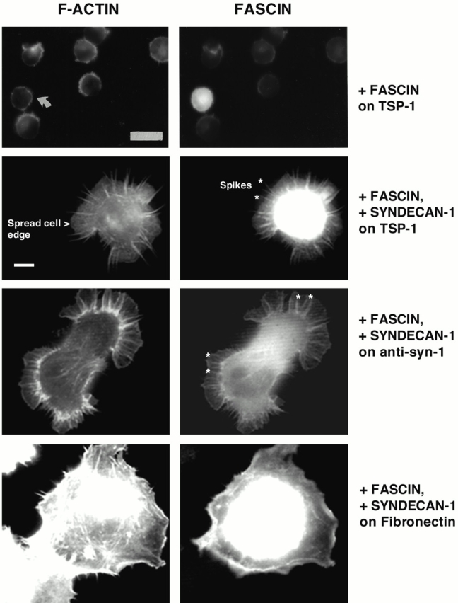
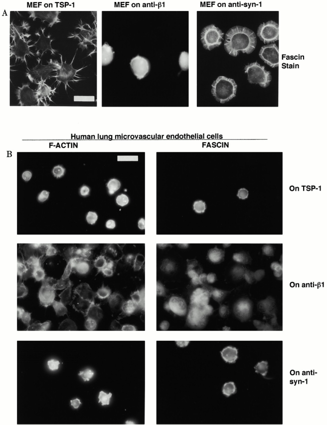
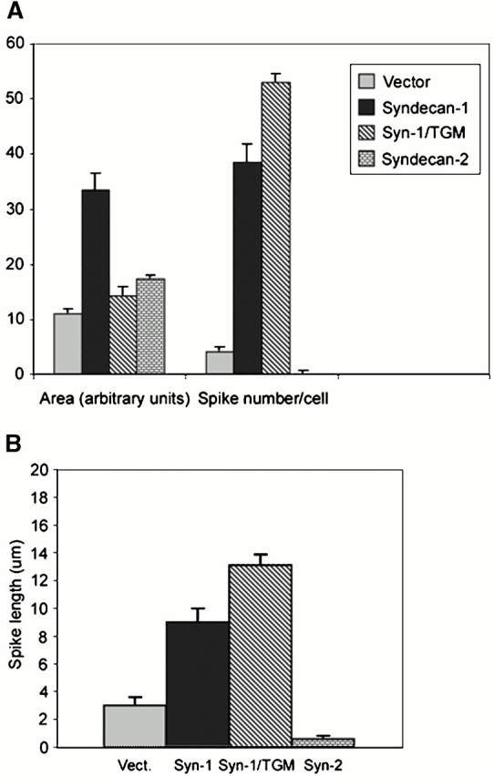
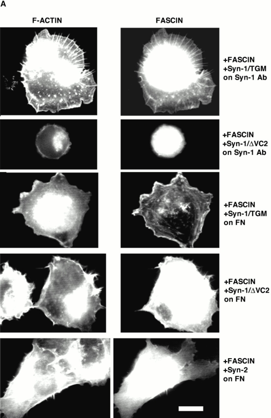
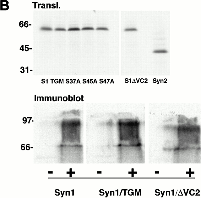
An important role of cell matrix adhesion receptors is to mediate transmembrane coupling between extracellular matrix attachment, actin reorganization, and cell spreading. Thrombospondin (TSP)-1 is a modulatory component of matrix expressed during development, immune response, or wound repair. Cell adhesion to TSP-1 involves formation of biochemically distinct matrix contacts based on stable fascin spikes. The cell surface adhesion receptors required have not been identified. We report here that antibody clustering of syndecan-1 proteoglycan specifically transduces organization of cortical actin and fascin bundles in several cell types. Transfection of COS-7 cells with syndecan-1 is sufficient to stimulate cell spreading, fascin spike assembly, and extensive protrusive lateral ruffling on TSP-1 or on syndecan-1 antibody. The underlying molecular mechanism depends on glycosaminoglycan (GAG) modification of the syndecan-1 core protein at residues S45 or S47 for cell membrane spreading and on the VC2 region of the cytoplasmic domain for spreading and fascin spike formation. Expression of the VC2 deletion mutant or GAG-negative syndecan-1 showed that syndecan-1 is necessary in spreading and fascin spike formation by C2C12 cells on TSP-1. These results establish a novel role for syndecan-1 protein in coupling a physiological matrix ligand to formation of a specific matrix contact structure.


Figures

Figure 1
Figure 1
Expression of TSP-1–binding receptors by C2C12 cells. (A) FACS® profiles. C2C12 cells were disaggregated with EDTA or with trypsin and stained with antibodies to the indicated adhesion receptors or nonimmune (NI) rat immunoglobulin. Similar profiles were obtained for EDTA (E) or trypsin-disaggregated (T) cells with the CD47, β1, and β3 antibodies (both profiles shown for β1 integrin only), whereas cells trypsinized for 2 min showed more heterogeneous syndecan-1 staining. (B) Syndecan-1 is expressed on C2C12 cells as a mixed proteoglycan. Proteoglycan extracts were prepared from C2C12 cells, resolved on gradient polyacrylamide gels under reducing conditions, transferred to nitrocellulose, and probed with antibody 281-2 to mouse syndecan-1. Lane 1, undigested extract; lane 2, + chondroitinase ABC digestion; lane 3, + heparitinase II digestion; lane 4, + digestion with both enzymes. Markers are indicated in kD. Results are representative of three experiments.
Figure 1
Figure 1
Expression of TSP-1–binding receptors by C2C12 cells. (A) FACS® profiles. C2C12 cells were disaggregated with EDTA or with trypsin and stained with antibodies to the indicated adhesion receptors or nonimmune (NI) rat immunoglobulin. Similar profiles were obtained for EDTA (E) or trypsin-disaggregated (T) cells with the CD47, β1, and β3 antibodies (both profiles shown for β1 integrin only), whereas cells trypsinized for 2 min showed more heterogeneous syndecan-1 staining. (B) Syndecan-1 is expressed on C2C12 cells as a mixed proteoglycan. Proteoglycan extracts were prepared from C2C12 cells, resolved on gradient polyacrylamide gels under reducing conditions, transferred to nitrocellulose, and probed with antibody 281-2 to mouse syndecan-1. Lane 1, undigested extract; lane 2, + chondroitinase ABC digestion; lane 3, + heparitinase II digestion; lane 4, + digestion with both enzymes. Markers are indicated in kD. Results are representative of three experiments.
Figure 2
Figure 2
F-actin organization in C2C12 cells adherent on adhesion receptor antibodies. Cells were plated onto surfaces, coated with 50 μg/ml of the indicated antibodies (see Materials and Methods) for 1 h in serum-free medium, then fixed and stained with TRITC-phalloidin. Bar, 15 μm.
Figure 3
Figure 3
Organization of fascin and vinculin in C2C12 cells adherent on adhesion receptor antibodies. (A) Cells were plated on surfaces coated with 50 μg/ml of the indicated anti–mouse antibodies for 1 h in serum-free medium and then fixed and stained for fascin or vinculin. (B) Fascin spike formation by primary mouse skeletal myoblasts on TSP-1. Wild-type or CD47-null skeletal myoblasts were plated on 50 nM TSP-1 for 1 h and then stained for fascin. Both populations formed extensive fascin spikes. Bar: (A, panels 1–6) 15 μm; (A, vinculin-stained panel) 8 μm; (B) 10 μm.
Figure 5
Figure 5
Expression of syndecan-1 specifically promotes cell spreading and formation of fascin spikes in TSP-1–adherent COS-7 cells. COS-7 cells were transfected with syndecan-1 and EGFP-fascin expression plasmids for 48 h. EDTA-released cells were plated on 50 nM TSP-1, fibro nectin, or 40 μg/ml anti–mouse syndecan-1 antibody in serum-free medium for 1 h and then fixed and stained for F-actin. Results shown are representative of three independent experiments, and ≥100 transfected cells were scored per experiment. The morphological features used for quantitation are also indicated. These were cell area measured by spread cell edge, and numbers and lengths of the radial fascin–actin bundles that form spikes and ribs. Examples of these bundles are marked with asteriks in two of the panels. Bars: (top two panels) 16 μm; (bottom six panels) 5 μm.
Figure 4
Figure 4
Organization of F-actin and fascin in cells adherent on TSP-1 or adhesion receptor antibodies. (A) EDTA-released mouse embryonic fibroblast cells (MEF) were plated on 50 nM TSP-1 or surfaces coated with 50 μg/ ml of the indicated antibodies as used in Fig. 1 and Fig. 2 for 1 h in serum-free medium and then fixed and stained for fascin. (B) EDTA-released HLMECs were plated on 50 nM TSP-1 or surfaces coated with 50 μg/ ml of antibodies to human adhesion receptors for 1 h in serum-free medium and then fixed and stained for F-actin or fascin. Bars, 20 μm.
Figure 6
Figure 6
GAG modifications of syndecan-1 and regions of the cytoplasmic domain are required for fascin spike formation on TSP-1. COS-7 cells were transfected with the indicated syndecan-1 mutants and EGFP-fascin expression plasmids, and 48 h later EDTA-released cells were plated on 50 nM TSP-1 in serum-free medium for 1 h and then fixed and stained for F-actin. Results shown are representative of three experiments, and ≥100 transfected cells were scored per experiment. Bar, 16 μm.
Figure 7
Figure 7
Expression of wild-type syndecan-1 promotes cell spreading and fascin spike assembly in TSP-1–adherent COS-7 cells. COS-7 cells transfected with empty expression vector or syndecan constructs were plated on 50 nM TSP-1 for 40 min in serum-free medium and then tracked by time-lapse videomicroscopy for 1 h. Cell areas and numbers of spikes (A) or spike lengths (B) were calculated from traces of spread cell margins after 1 h of adhesion to TSP-1. See Fig. 5 for examples of the morphological features. Bars show mean ± SEM for data from three experiments.
Figure 8
Figure 8
The effects of GAG removal from syndecan-1 are specific to TSP-1 adhesion. (A) COS-7 cells transfected for 48 h with the indicated syndecan constructs and EGFP-fascin were EDTA-released for 1-h adhesion assays on antibody to mouse syndecan-1 or 50 nM fibronectin. Actin organization was compared with EGFP-fascin distribution in the transfected cells. Results shown are representative of three experiments, and ≥50 transfected cells were scored per experiment. (B) Relative expression levels of syndecans used in this study. Top, in vitro translation of 1 μg of each syndecan construct detected by [35S]methionine incorporation; bottom, immunoblot of double heparitinase and chondroitinase–digested syndecan-1 proteins after expression and extraction from COS-7 cells detected by 281-2 antibody. Molecular weight markers are given in kD. Bar, 10 μm.
Figure 8
Figure 8
The effects of GAG removal from syndecan-1 are specific to TSP-1 adhesion. (A) COS-7 cells transfected for 48 h with the indicated syndecan constructs and EGFP-fascin were EDTA-released for 1-h adhesion assays on antibody to mouse syndecan-1 or 50 nM fibronectin. Actin organization was compared with EGFP-fascin distribution in the transfected cells. Results shown are representative of three experiments, and ≥50 transfected cells were scored per experiment. (B) Relative expression levels of syndecans used in this study. Top, in vitro translation of 1 μg of each syndecan construct detected by [35S]methionine incorporation; bottom, immunoblot of double heparitinase and chondroitinase–digested syndecan-1 proteins after expression and extraction from COS-7 cells detected by 281-2 antibody. Molecular weight markers are given in kD. Bar, 10 μm.
Figure 9
Figure 9
Role of endogenous syndecan-1 in fascin spike formation by C2C12 cells adherent on TSP-1. In a separate protocol, C2C12 cotransfected with Syn-1/ΔVC2 or Syn-1/TGM and EGFP-fascin expression plasmids were identified by EGFP-fascin expression after 1 h adhesion on TSP-1. Bar, 12 μm.
Figure 10
Figure 10
Effects of syndecan expression on aspects of cell motile behavior. Apical projections and lateral protrusive ruffling activity of cells adherent on 50 nM TSP-1 were scored from phase–contrast time-lapse videos that tracked vector or syndecan transfectant cells from 45 min to 1.5 h of adhesion. Locomotion was scored as the displacement of cell centroids between 45 min and 2 h of adhesion. Bars show mean ± SEM for data from five experiments. Shade code same as in Fig. 7.

References

    1. Adams J.C. Formation of stable microspikes containing actin and the 55 kDa actin bundling protein, fascin, is a consequence of cell adhesion to thrombospondin-1implications for the anti-adhesive activities of thrombospondin-1. J. Cell Sci. 1995;108:1977–1990. - PubMed
    1. Adams J.C. Thrombospondin-1 Int. J. Biochem. Cell Biol 29 1997. 861 865a - PubMed
    1. Adams J.C. Characterization of cell-matrix adhesion requirements for the formation of fascin microspikes Mol. Biol. Cell. 8 1997. 2345 2363b - PMC - PubMed
    1. Adams J.C., Lawler J. Diverse mechanisms for cell attachment to platelet thrombospondin. J. Cell Sci. 1993;104:1061–1071. - PubMed
    1. Adams J.C., Lawler J. Cell-type specific adhesive interactions of skeletal myoblasts with thrombospondin-1. Mol. Biol. Cell. 1994;5:623–637. - PMC - PubMed

Publication types

MeSH terms

Substances