. 2001 Mar 19;152(6):1219-32.
doi: 10.1083/jcb.152.6.1219.

The NC1/endostatin domain of Caenorhabditis elegans type XVIII collagen affects cell migration and axon guidance

Affiliations

The NC1/endostatin domain of Caenorhabditis elegans type XVIII collagen affects cell migration and axon guidance

B D Ackley et al. J Cell Biol. .

Abstract

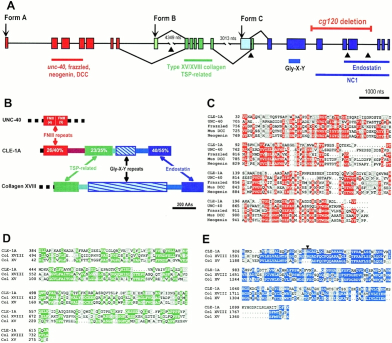
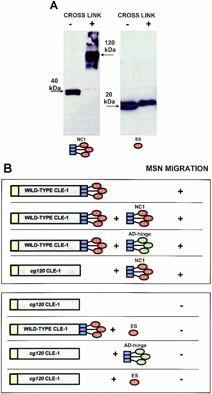
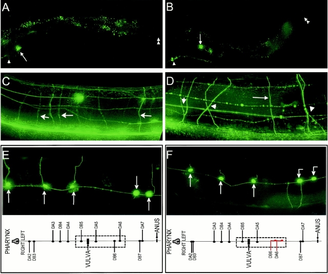
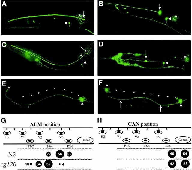
Type XVIII collagen is a homotrimeric basement membrane molecule of unknown function, whose COOH-terminal NC1 domain contains endostatin (ES), a potent antiangiogenic agent. The Caenorhabditis elegans collagen XVIII homologue, cle-1, encodes three developmentally regulated protein isoforms expressed predominantly in neurons. The CLE-1 protein is found in low amounts in all basement membranes but accumulates at high levels in the nervous system. Deletion of the cle-1 NC1 domain results in viable fertile animals that display multiple cell migration and axon guidance defects. Particular defects can be rescued by ectopic expression of the NC1 domain, which is shown to be capable of forming trimers. In contrast, expression of monomeric ES does not rescue but dominantly causes cell and axon migration defects that phenocopy the NC1 deletion, suggesting that ES inhibits the promigratory activity of the NC1 domain. These results indicate that the cle-1 NC1/ES domain regulates cell and axon migrations in C. elegans.


Figures

Figure 1
Figure 1
cle-1 Gene and protein structures. (A) Genomic structure of cle-1 (sequence data available from GenBank/EMBL/DDBJ under accession number AF164959). Exons are indicated as boxes; introns, as lines. Form A–specific exons are red, exons common to forms A and B are dark green, and exons common to all forms are dark blue. The predicted translation start site of each isoform is indicated by an arrow. Splicing patterns for inclusion of the form B– or C–specific first exons (light green or light blue, respectively) are indicated above the intron/exon structure, and splicing patterns for the longer forms, lacking these exons, are below. The four introns that are conserved in cle-1 and mammalian type XVIII and XV collagens are marked with arrowheads. Extents of CLE-1 protein domains are indicated below the structure, and extents of the cg120 deletion are indicated above. (B) CLE-1A domain structure and relatedness to mouse type XVIII collagen and C. elegans UNC-40. Homologous domains are indicated with the same colors. Percentages of amino acid sequence identity and similarity are indicated in the relevant CLE-1A domains (identity/similarity). The regions connecting the thrombospondin (TSP)-related and ES domains to the Gly-X-Y repeats are 34% similar between CLE-1 and type XVIII collagen. (C) Alignment of CLE-1A fibronectin type III (FNIII) repeats with C. elegans UNC-40 (AAB17088), Drosophila Frazzled (AAC47314.1), mouse DCC (P70211), and mouse neogenin (CAA70727.1). (D) Alignment of CLE-1A TSP-related motifs with mouse collagen types XVIII (AAC52903) and XV (AAC53387). (E) Alignment of the CLE-1A COOH-terminal domain with mouse ES (Col XVIII) and restin (Col XV). The four amino acid loop containing Arg158 (arrowhead) is present in CLE-1 and type XVIII collagen but not type XV collagen.
Figure 2
Figure 2
Expression patterns of cle-1::GFP transcriptional reporters. Isoform-specific GFP transcriptional reporters show the expression patterns of cle-1A (A and E), cle-1B (B and F), and cle-1C (C, D, G, and H). In A–D, anterior is up and dorsal is to the left; in E–H, anterior is to the left, and dorsal is up. (A) cle-1A expression in the cephalic neurons of a 1.75-fold stage embryo. Expression is also seen in S-type interneurons at later stages. (B) cle-1B expression in a threefold embryo is seen in DD ventral motorneurons (four are visible and two are out of the plane of focus). This GFP fusion localizes to the nucleus, making axon visualization difficult. (C) cle-1C expression in the body wall muscles (bm) of a twofold embryo. Pharyngeal expression is not seen in this focal plane. (D) In a threefold embryo, cle-1C expression in body wall muscles is weaker, but strong expression is seen in pharyngeal cells (p), accessory muscles, some unidentified cells near the anus (a), and the canal-associated neurons (CAN). (E) In an L2 larva, cle-1A expression is strong in the nerve ring (nr) and rectal epithelial cells (re). GFP is weakly detected in axons of the S-type interneurons that run sublaterally. (F) cle-1B expression in dorsal motorneuron cell bodies (arrowheads) located along the ventral nerve cord of an L2 larva. Faint GFP activity can be detected in commissural axons of most animals. (G) cle-1C expression in an L2 larva is limited to a subset of glial-like GLR cells, the head mesodermal cell (hm), the canal-associated neurons, and weakly in the anal sphincter muscle (sm). (H) cle-1C expression in adult body wall muscles (bm), accessory muscles (sm), canal associated neurons (CAN), and gonadal sheath cells (sc).
Figure 3
Figure 3
Localization of CLE-1 and type IV collagen in wild-type and cg120 animals. Immunofluorescent localization of CLE-1 (A–H) and type IV collagen (I and J) are shown in wild-type (A–D and I) and cg120 (E–H and J) animals. In all panels, anterior is left, and dorsal is up. (A) A fourfold embryo shows strong CLE-1 staining associated with the nerve ring (arrowhead). (B) In larval animals, CLE-1 accumulates on the nerve ring (arrowhead) and the dorsal (dc) and ventral (vc) nerve cords. (C) In adults, staining for CLE-1 is strong on the nerve ring and nerve cords (dc and vc) and is weakly detected on the surfaces of the pharynx (p), intestine (i), and gonad (g). (D) Under body wall muscle of an adult, CLE-1 accumulates strongly at the junctions between muscle cells (arrowheads) and along muscle-dense body lines (small arrows) but is not coincident with the dense bodies. Staining on the ventral nerve cord is distinctly punctate. (E) Weak CLE-1 accumulation on the nerve ring of a cg120 embryo. (F) CLE-1 localization to the nerve ring (arrowhead) and dorsal (dc) and ventral (vc) nerve cords of a cg120 larva. (G) Accumulation on nerve ring (arrowhead), nerve cords (dc and vc), pharynx (p), and intestine (i) of a cg120 adult. (H) Under the body wall muscle of a cg120 adult, CLE-1 accumulates along dense body lines (small arrows) and along the ventral nerve cord (vc) as in wild type. However, there is no accumulation at the junctions between muscle cells (arrowheads), and the pattern on the nerve cord is more diffuse. (I and J) Wild-type and cg120 L1 larvae show type IV collagen localization in the basement membranes of the pharynx (p), intestine (i), and gonad primordium (g).
Figure 8
Figure 8
The C. elegans NC1 domain trimerizes in vitro. (A) Purified CLE-1 NC1 (left) and ES domains (right) were analyzed by denaturing gel electrophoresis and Western blotting, either before (−) or after (+) cross-linking with EGS. After cross-linking, the 40-kD NC1 monomer migrates at ∼120 kD, indicating that it exists as a trimer. The mobility of the ES domain does not shift after EGS treatment, indicating that it exists as a monomer. (B) Summary of mechanosensory neuron migration data. Defective mechanosensory neuron migrations are observed when the NC1 domain is removed by the cg120 deletion. These defects can be rescued by ectopic NC1 domain expression. In contrast, ectopic ES domain expression causes mechanosensory neuron migration defects in the wild-type background and fails to rescue the NC1 deletion defects. Expression of the association domain and hinge regions fused to GFP, rather than ES, does not rescue cg120 migration defects and does not cause defects in the wild-type background.
Figure 4
Figure 4
Cell and axon migration defects in cle-1(cg120) animals. Cell and axon positions were analyzed in wild-type (A, C, and E) and cg120 mutant (B, D, and F) animals. Anterior is left, and dorsal is up in all panels. (A and B) HSNs visualized using the tph-1::GFP marker (Sze et al. 2000) are indicated with an arrow. The positions of the vulva (arrowhead) and anus (double arrowhead) are marked to visualize the relative position of the HSN. (A) In wild-type animals, the left HSN is positioned immediately posterior of the vulva. (B) In this cg120 animal, the HSN is displaced posteriorly and dorsally. The HSN axon abnormally projects posteriorly and then ventrally to the ventral cord. (C and D) A panneuronal marker (F25B3.3::GFP) was used to visualize the general organization of the nervous system. (C) In wild-type animals, dorsoventral axon migrations (arrows) follow a trajectory that is orthogonal to the anterior–posterior axis. (D) Axons in cg120 mutants frequently follow nonorthogonal trajectories and defasciculate from one another (arrowheads), but some axon trajectories are normal (arrow). (E and F) Visualization of DA and DB motorneurons using the unc129::GFP reporter. Schematics of the complete DA and DB cell body and axon patterns are shown below, and the area shown in the micrographs is boxed with a dashed line. (E) In wild-type animals, the DA and DB motorneurons extend commissural axons directly from their cell bodies. (F) In this cg120 animal, the DA6 and DB6 axons abnormally migrate anteriorly along the ventral cord, and then both exit on the right side. The DA6 axon should exit on the left side of the ventral cord.
Figure 5
Figure 5
Cell positioning and axon guidance of mechanosensory neurons. The mec-7::GFP marker was used to visualize the anterior (A–D) or posterior (E–F) mechanosensory neuron cell bodies and axons. Anterior is left, and dorsal is up in all panels. (A) In wild-type animals, ALMR (arrow) is located posterior and dorsal of AVM (arrowhead). ALML (*) is out of the plane of focus. (B) In a cg120 mutant, ALMR (arrow) is located anterior of AVM (arrowhead), whereas ALML (*) is normally positioned. (C) In a cg120 animal expressing mec-7::CelNC1, the anterior mechanosensory neurons are normally positioned. (D) In a wild-type animal expressing mec-7::CelES, ALMR (arrow) is located both anterior and ventral of its normal position, whereas ALML is normally positioned. (E and F) PLM cell bodies are not mispositioned in cg120, so their axons were analyzed for guidance defects. (E) In wild-type animals, the PLMR axon (large arrowhead) extends anteriorly along the sublateral tract and terminates just posterior of the ALMR cell body (*). The dorsal edge of the animal is indicated (small arrowheads). (F) In a cg120 animal, the PLMR axon deviates dorsally from the normal ventral sublateral position over a segment of its path (arrows). (G and H) The positions of ALM mechanosensory neurons (G) and canal-associated neurons (CAN; H) were scored in first larval stage animals immediately after hatching using differential interference contrast optics. Cell positions were scored relative to the hypodermal nuclei, which are represented by ovals in the upper part of the drawing. These nuclei are in fixed positions and are used as static markers to score cell positions. Numbers and circles in the lower part of the drawing indicate the percent of animals (n = 50) with cells in the indicated positions.
Figure 6
Figure 6
Male tail and gonad migration defects. Defects in morphogenesis of the male tail (A–C) and the hermaphrodite gonad (D and E), visualized with differential interference contrast microscopy. (A) Ventral view of a wild-type male tail showing the nine bilateral pairs of sensory rays, labeled 1–9, on the left side of the animal. (B) A cg120 male tail shows fusion of rays 8 and 9 (8,9). On the right side, ray 6 is broader than normal, and the cuticle covering it is irregular. (C) The tail of a male ectopically expressing mec-7::CelES shows fusion of rays 1 and 2 (1,2) on both sides of the animal. Ray 6 on the right side (6) appears crumpled, and rays 8 and 9 on the left have fused (8,9). The fans and rays of males expressing mec-7::CelES are smaller than those of wild type. (D) The anterior arm of a wild-type fourth larval stage hermaphrodite gonad. The distal tip cell (DTC) leads gonad migration anteriorly along the dorsal body wall, turns and migrates to the dorsal side, and then migrates posteriorly. The position of the vulva (V) is indicated. A schematic depiction of the DTC migration path is shown to the right of the micrograph. 100% (50/50) of wild-type animals showed this migration pattern. (E) In this cg120 animal, the DTC migrated anteriorly, turned dorsal prematurely while continuing to migrate anteriorly, and then reflexed and migrated posteriorly (dashed line) along the dorsal body wall. The final posterior migration is below the plane of focus. 32% (16/50) of cg120 animals displayed similar migration defects of the anterior gonad. 16% of posterior gonads showed the same defects.
Figure 7
Figure 7
RNAi against cle-1 results in embryonic and larval lethality. (A–D) Embryonic lethality resulting from RNAi against cle-1. Embryos are oriented with anterior up and ventral to the right. The position of the presumptive oral cavity is indicated by an arrowhead. (A) A wild-type embryo at the 1.5-fold stage of embryogenesis. (B) An embryo arrested at the 1.5–2-fold stage of embryogenesis resulting from RNAi into the wild-type background. The embryo has herniated at the ventral pocket (arrow), which forms during enclosure by the hypodermis. (C and D) Embryos arrested at the 1.5-fold stage of embryogenesis resulting from RNAi into the cg120 background. The embryos are small and misshapen, suggesting defects in hypodermal function. (E and F) Larval lethality resulting from RNAi against cle-1. The posterior bulb of the pharynx is indicated by an arrow, and the anterior bulb by an arrowhead. (E) Wild-type first stage larva. The body has a uniform diameter along its length, and the pharynx lies along the central axis of the animal. (F) Arrested first stage larva resulting from RNAi into the wild type background. The animal is smaller than wild type, and the posterior body is shrunken relative to the anterior. The pharynx is mislocalized laterally and does not pump.

References

    1. Barstead R.J. Reverse genetics. In: Hope I.A., editor. C. elegansA Practical Approach. Oxford University Press; Oxford, UK: 1999. pp. 97–118.
    1. Baum P.D., Garriga G. Neuronal migrations and axon fasciculation are disrupted in ina-1 integrin mutants. Neuron. 1997;19:51–62. - PubMed
    1. Bettinger J.C., Lee K., Rougvie A.E. Stage-specific accumulation of the terminal differentiation factor LIN-29 during Caenorhabditis elegans development. Development. 1996;122:2517–2527. - PubMed
    1. Boehm T., Folkman J., Browder T., O'Reilly M.S. Antiangiogenic therapy of experimental cancer does not induce acquired drug resistance. Nature. 1997;390:404–407. - PubMed
    1. Brenner S. The genetics of Caenorhabditis elegans . Genetics. 1974;77:71–94. - PMC - PubMed

Publication types

MeSH terms