. 2001 Mar 19;152(6):1233-46.
doi: 10.1083/jcb.152.6.1233.

Oligomerization-dependent regulation of motility and morphogenesis by the collagen XVIII NC1/endostatin domain

Affiliations

Oligomerization-dependent regulation of motility and morphogenesis by the collagen XVIII NC1/endostatin domain

C J Kuo et al. J Cell Biol. .

Abstract

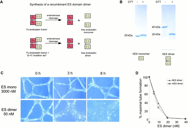
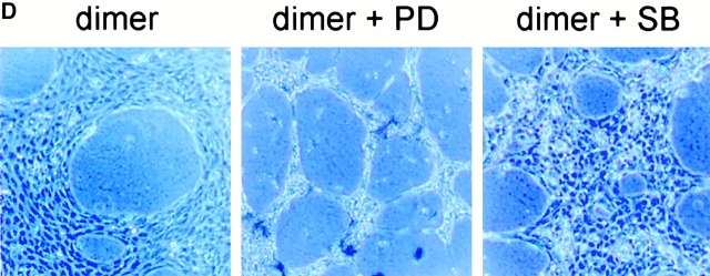
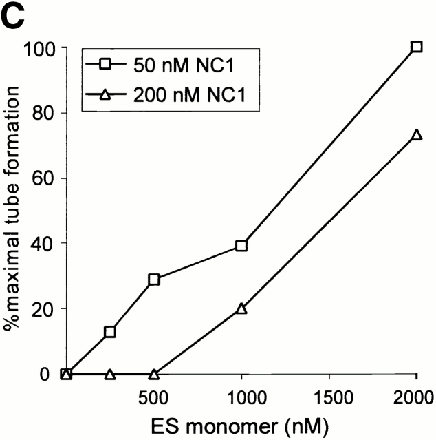
Collagen XVIII (c18) is a triple helical endothelial/epithelial basement membrane protein whose noncollagenous (NC)1 region trimerizes a COOH-terminal endostatin (ES) domain conserved in vertebrates, Caenorhabditis elegans and Drosophila. Here, the c18 NC1 domain functioned as a motility-inducing factor regulating the extracellular matrix (ECM)-dependent morphogenesis of endothelial and other cell types. This motogenic activity required ES domain oligomerization, was dependent on rac, cdc42, and mitogen-activated protein kinase, and exhibited functional distinction from the archetypal motogenic scatter factors hepatocyte growth factor and macrophage stimulatory protein. The motility-inducing and mitogen-activated protein kinase-stimulating activities of c18 NC1 were blocked by its physiologic cleavage product ES monomer, consistent with a proteolysis-dependent negative feedback mechanism. These data indicate that the collagen XVIII NC1 region encodes a motogen strictly requiring ES domain oligomerization and suggest a previously unsuspected mechanism for ECM regulation of motility and morphogenesis.


Figures

Figure 1
Figure 1
Inhibition of in vitro endothelial tube assembly by collagen XVIII NC1. (A) Topology of collagen XVIII. Signal peptide, triple helical collagenous repeats, NC1 domain, and a protease-sensitive “hinge” region interposed within the trimerization and ES motifs are indicated. Dotted line indicates alternative splice which occurs in nonhepatic tissues and removes the frizzled homology domain. (B) Production of recombinant c18 NC1. Human c18 NC1 purified from supernatant of stably transfected 293T cells was analyzed by SDS-PAGE with or without EGS cross-linking. Oligomeric forms corresponding to NC1 trimer at ∼114 kD (38 × 3) and 76-kD species from incomplete cross-linking (38 × 2) are indicated. (C) Recombinant c18 NC1 inhibits in vitro endothelial tube formation. HUVECs were plated on Matrigel in the absence (untreated) or presence of murine or human c18 NC1 at 50 nM and photographed under phase–contrast after 16 h (magnification ×40). A complete lack of tubular structures and a dispersed cell phenotype are apparent in the NC1-treated wells. (D) Dose-dependent inhibition of endothelial tube formation by recombinant c18 NC1. Indicated amounts of human or mouse c18 NC1 were added at the time of HUVEC seeding onto Matrigel-covered wells. Tubular structures were quantitated by manual counting of low power fields after 16 h and percent inhibition was expressed using untreated wells as 100%.
Figure 1
Figure 1
Inhibition of in vitro endothelial tube assembly by collagen XVIII NC1. (A) Topology of collagen XVIII. Signal peptide, triple helical collagenous repeats, NC1 domain, and a protease-sensitive “hinge” region interposed within the trimerization and ES motifs are indicated. Dotted line indicates alternative splice which occurs in nonhepatic tissues and removes the frizzled homology domain. (B) Production of recombinant c18 NC1. Human c18 NC1 purified from supernatant of stably transfected 293T cells was analyzed by SDS-PAGE with or without EGS cross-linking. Oligomeric forms corresponding to NC1 trimer at ∼114 kD (38 × 3) and 76-kD species from incomplete cross-linking (38 × 2) are indicated. (C) Recombinant c18 NC1 inhibits in vitro endothelial tube formation. HUVECs were plated on Matrigel in the absence (untreated) or presence of murine or human c18 NC1 at 50 nM and photographed under phase–contrast after 16 h (magnification ×40). A complete lack of tubular structures and a dispersed cell phenotype are apparent in the NC1-treated wells. (D) Dose-dependent inhibition of endothelial tube formation by recombinant c18 NC1. Indicated amounts of human or mouse c18 NC1 were added at the time of HUVEC seeding onto Matrigel-covered wells. Tubular structures were quantitated by manual counting of low power fields after 16 h and percent inhibition was expressed using untreated wells as 100%.
Figure 4
Figure 4
Oligomerized collagen XV ES domains do not exhibit motogenic activity. HUVECs were plated on Matrigel in the presence of mNC1(c18) (50 nM), mNC1(c15) (100 nM), or Fc-mES(c15) (250 nM) and photographed after 20 h. Identical results were obtained upon application of factors to preformed tubules.
Figure 2
Figure 2
Stimulation of endothelial cell motility by collagen XVIII NC1. (A) Rapid induction of motility by human c18 NC1. HUVECs were plated on Matrigel for 16 h and allowed to form tubular structures, followed by recombinant human c18 NC1 treatment (50 nM) for 2 h. Top, phase–contrast microscopy (magnification ×200); bottom, confocal microscopy. Cells treated as above were stained with phalloidin-FITC to stain filamentous actin and photographed under confocal microscopy. NC1-treated cells exhibit prominent cell migration with formation of stress fibers, filopodia, and lamellipodia. (B) Migration of HUVECs from preestablished tubules is stimulated by c18 NC1. HUVECs were plated on Matrigel for 16 h and identical fields were photographed under phase–contrast before (0 h) and after (8 h) stimulation by recombinant human c18 NC1 (50 nM) (magnification ×40).
Figure 3
Figure 3
ES domain oligomerization is necessary and sufficient for c18-induced motility. (A) Schematic of synthesis of ES domain monomer and dimer. Cleavage of an Fc-ES domain fusion produces free ES monomer. In contrast, cleavage of Fc-ES(Q7→C7) produces an ES domain dimer because of disulfide bond formation between adjacent C7 residues. (B) Nonreducing and reducing SDS-PAGE of purified recombinant human ES monomer and dimer. Migration shift from 40 to 20 kD is noted for ES dimer but not ES monomer upon disulfide reduction with DTT. (C) ES dimer but not ES monomer stimulates migration of HUVECs from pre-formed tubules on Matrigel. HUVECs were plated on Matrigel and allowed to form tubes for 16 h, followed by stimulation with ES monomer (3,000 nM) or ES dimer (50 nM) for the indicated times. Representative fields were photographed under phase–contrast (magnification ×200). (D) Dose response to recombinant human ES dimer. HUVECs were plated on Matrigel in the presence of hES dimer, and after 16 h tubular structures were quantitated by manual counting of central fields and percentage of maximal tube formation was calculated. (E) Induction of motility by Fc-ES fusions which dimerize the ES domain. Preformed HUVEC tubules were treated with Fc-mES(c18) (40 nM), Fc (3,000 nM), or mES(c18) (3,000 nM) and photographed after 30 h (magnification ×40). (F) Summary of motogenic activity of monomeric, dimeric, and trimeric ES domain derivatives.
Figure 3
Figure 3
ES domain oligomerization is necessary and sufficient for c18-induced motility. (A) Schematic of synthesis of ES domain monomer and dimer. Cleavage of an Fc-ES domain fusion produces free ES monomer. In contrast, cleavage of Fc-ES(Q7→C7) produces an ES domain dimer because of disulfide bond formation between adjacent C7 residues. (B) Nonreducing and reducing SDS-PAGE of purified recombinant human ES monomer and dimer. Migration shift from 40 to 20 kD is noted for ES dimer but not ES monomer upon disulfide reduction with DTT. (C) ES dimer but not ES monomer stimulates migration of HUVECs from pre-formed tubules on Matrigel. HUVECs were plated on Matrigel and allowed to form tubes for 16 h, followed by stimulation with ES monomer (3,000 nM) or ES dimer (50 nM) for the indicated times. Representative fields were photographed under phase–contrast (magnification ×200). (D) Dose response to recombinant human ES dimer. HUVECs were plated on Matrigel in the presence of hES dimer, and after 16 h tubular structures were quantitated by manual counting of central fields and percentage of maximal tube formation was calculated. (E) Induction of motility by Fc-ES fusions which dimerize the ES domain. Preformed HUVEC tubules were treated with Fc-mES(c18) (40 nM), Fc (3,000 nM), or mES(c18) (3,000 nM) and photographed after 30 h (magnification ×40). (F) Summary of motogenic activity of monomeric, dimeric, and trimeric ES domain derivatives.
Figure 5
Figure 5
Motile response of nonendothelial cell types to c18 derivatives. PC12 and 293T cells were cultured with hES monomer (3,000 nM), hES dimer (25 nM), or hNC1 (50 nM) on Matrigel for 16 h and photographed under phase–contrast (magnification ×200 [bottom]).
Figure 8
Figure 8
Motogenic activity of c18 NC1 requires rac and cdc42. (A) Inhibition of c18-stimulated motility by C. difficile toxin B. HUVECs on Matrigel for 12 h were preincubated with or without C. difficile toxin B (50 ng/ml) for 4 h and treated with hES monomer (3,000 nM), hES dimer (50 nM), or c18 NC1 (50 nM) for 16 h, then photographed under phase–contrast (magnification ×200). Trypan blue staining of these cultures revealed <5% toxicity from the toxin treatment. Cell-rounding characteristic of rho inhibition is present in +toxin panels. (B) Inhibition of c18 NC1-stimulated motility by adenovirus-encoded DN alleles of rac and cdc42. Top, HUVEC tubules previously infected with adenovirus encoding racDN, rhoDN, or cdc42DN were treated with hES dimer (75 nM) and photographed (magnification ×200). Strong inhibition of motility by racDN and cdc42DN is present. RhoDN was ineffective at blocking scatter, although cell-rounding characteristic of rho inhibition was observed. Bottom, Parallel cultures were treated identically and processed for immunofluorescence using anti-myc epitope tag (rac, cdc42) or anti-rho (rhoDN and dimer alone), revealing >95% infection with racDN, rhoDN, and cdc42DN.
Figure 8
Figure 8
Motogenic activity of c18 NC1 requires rac and cdc42. (A) Inhibition of c18-stimulated motility by C. difficile toxin B. HUVECs on Matrigel for 12 h were preincubated with or without C. difficile toxin B (50 ng/ml) for 4 h and treated with hES monomer (3,000 nM), hES dimer (50 nM), or c18 NC1 (50 nM) for 16 h, then photographed under phase–contrast (magnification ×200). Trypan blue staining of these cultures revealed <5% toxicity from the toxin treatment. Cell-rounding characteristic of rho inhibition is present in +toxin panels. (B) Inhibition of c18 NC1-stimulated motility by adenovirus-encoded DN alleles of rac and cdc42. Top, HUVEC tubules previously infected with adenovirus encoding racDN, rhoDN, or cdc42DN were treated with hES dimer (75 nM) and photographed (magnification ×200). Strong inhibition of motility by racDN and cdc42DN is present. RhoDN was ineffective at blocking scatter, although cell-rounding characteristic of rho inhibition was observed. Bottom, Parallel cultures were treated identically and processed for immunofluorescence using anti-myc epitope tag (rac, cdc42) or anti-rho (rhoDN and dimer alone), revealing >95% infection with racDN, rhoDN, and cdc42DN.
Figure 6
Figure 6
Comparison of c18 and HGF motogenic activities. HUVEC tubules on Matrigel for 18 h (top) or MDCK cells on plastic (bottom) were treated with HGF (50 ng/ml), hES monomer (3,500 nM), hES dimer (50 nM), or c18 NC1/hES trimer (50 nM) for 24 h and photographed. Top, magnification ×200; bottom, ×100. Note complete absence of HGF response in HUVECs on Matrigel and complete absence of c18 responses in MDCK cells on plastic.
Figure 7
Figure 7
Blockade of the MAPK pathway antagonizes c18-induced motility. (A) Stimulation of MAPK phosphorylation by c18 NC1 and ES dimer. HUVECs on Matrigel in the presence or absence of ES monomer (3,000 nM), ES dimer (50 nM), or c18 NC1 (50 nM), and harvested and analyzed by Western blotting using anti–phospho-MAPK antisera after 24 h. (B) Time kinetics of MAPK activation by ES dimer. HUVECs on Matrigel for 16 h were stimulated with ES dimer (50 nM) or ES monomer (3,000 nM) for the indicated times, followed by harvest and Western blotting with anti–phospho-MAPK antisera. Stimulation was measured by densitometry and represents the average of three independent experiments. Similar results were observed for NC1. (C and D) Inhibition of ES dimer–induced motility by the MEK inhibitor PD98056 but not the p38 inhibitor SB203580. HUVECs were seeded on Matrigel for 16 h followed by 60-min preincubation with or without PD98056 or SB203580 (50 μM), followed by treatment with hES dimer (50 nM) for 12 h, and then harvest for anti–phospho-MAPK Western blot (C) or phase–contrast microscopy (D) (magnification ×40).
Figure 7
Figure 7
Blockade of the MAPK pathway antagonizes c18-induced motility. (A) Stimulation of MAPK phosphorylation by c18 NC1 and ES dimer. HUVECs on Matrigel in the presence or absence of ES monomer (3,000 nM), ES dimer (50 nM), or c18 NC1 (50 nM), and harvested and analyzed by Western blotting using anti–phospho-MAPK antisera after 24 h. (B) Time kinetics of MAPK activation by ES dimer. HUVECs on Matrigel for 16 h were stimulated with ES dimer (50 nM) or ES monomer (3,000 nM) for the indicated times, followed by harvest and Western blotting with anti–phospho-MAPK antisera. Stimulation was measured by densitometry and represents the average of three independent experiments. Similar results were observed for NC1. (C and D) Inhibition of ES dimer–induced motility by the MEK inhibitor PD98056 but not the p38 inhibitor SB203580. HUVECs were seeded on Matrigel for 16 h followed by 60-min preincubation with or without PD98056 or SB203580 (50 μM), followed by treatment with hES dimer (50 nM) for 12 h, and then harvest for anti–phospho-MAPK Western blot (C) or phase–contrast microscopy (D) (magnification ×40).
Figure 9
Figure 9
Autoregulatory inhibition of c18 NC1 by the cleavage product ES monomer. (A) ES monomer inhibits motility induced by c18 NC1 or hES dimer. Left panels, HUVEC tubules on Matrigel for 12 h were treated with ES dimer (50 nM) or NC1/ES trimer (75 nM) for 24 h after 30-min preincubation with human ES monomer (3,000 nM) as appropriate, followed by phase–contrast microscopy (magnification ×40). B, PC12 cells were plated on Matrigel in the presence of ES dimer (50 nM) or NC1/ES trimer (75 nM) for 20 h with or without 30-min preincubation with human ES monomer (3,000 nM) as appropriate, followed by phase–contrast microscopy (magnification ×200). Similar results were obtained with treatment with factors after 12–16-h culture on Matrigel. (C) Dose-dependent reversal of c18 NC1 inhibition of HUVEC tube formation by ES monomer. Increasing concentrations of hES monomer were added to freshly seeded HUVECs on Matrigel, followed after 30 min by addition of c18 NC1 (50 or 200 nM) and quantitation of tube formation after 16 h. Higher concentrations of ES monomer were required to reverse inhibition by 200 nM as opposed to 50 nM NC1. (D) Stimulation of MAPK by c18 NC1 or ES dimer is inhibited by ES monomer. HUVEC tubules on Matrigel for 16 h were preincubated with 3,000 nM hES monomer or PBS for 30 min as appropriate, followed by stimulation with hNC1 or hES dimer (50 nM) for 24 h and Western blotting with anti–phospho- MAPK antibody.
Figure 9
Figure 9
Autoregulatory inhibition of c18 NC1 by the cleavage product ES monomer. (A) ES monomer inhibits motility induced by c18 NC1 or hES dimer. Left panels, HUVEC tubules on Matrigel for 12 h were treated with ES dimer (50 nM) or NC1/ES trimer (75 nM) for 24 h after 30-min preincubation with human ES monomer (3,000 nM) as appropriate, followed by phase–contrast microscopy (magnification ×40). B, PC12 cells were plated on Matrigel in the presence of ES dimer (50 nM) or NC1/ES trimer (75 nM) for 20 h with or without 30-min preincubation with human ES monomer (3,000 nM) as appropriate, followed by phase–contrast microscopy (magnification ×200). Similar results were obtained with treatment with factors after 12–16-h culture on Matrigel. (C) Dose-dependent reversal of c18 NC1 inhibition of HUVEC tube formation by ES monomer. Increasing concentrations of hES monomer were added to freshly seeded HUVECs on Matrigel, followed after 30 min by addition of c18 NC1 (50 or 200 nM) and quantitation of tube formation after 16 h. Higher concentrations of ES monomer were required to reverse inhibition by 200 nM as opposed to 50 nM NC1. (D) Stimulation of MAPK by c18 NC1 or ES dimer is inhibited by ES monomer. HUVEC tubules on Matrigel for 16 h were preincubated with 3,000 nM hES monomer or PBS for 30 min as appropriate, followed by stimulation with hNC1 or hES dimer (50 nM) for 24 h and Western blotting with anti–phospho- MAPK antibody.

References

    1. Ackley B.D., Crew J.R., Elamaa H., Pihlajaniemi T., Kuo C.J., Kramer J.M. The NC1/endostatin domain of Caenorhabditis elegans type XVIII collagen affects cell migration and axon guidance. J. Cell Biol. 2001;152:1219–1232. - PMC - PubMed
    1. Binari R.C., Staveley B.E., Johnson W.A., Godavarti R., Sasisekharan R., Manoukian A.S. Genetic evidence that heparin-like glycosaminoglycans are involved in wingless signaling. Development. 1997;124:2623–2632. - PubMed
    1. Bussolino F., Di Renzo M.F., Ziche M., Bocchietto E., Olivero M., Naldini L., Gaudino G., Tamagnone L., Coffer A., Comoglio P.M. Hepatocyte growth factor is a potent angiogenic factor which stimulates endothelial cell motility and growth. J. Cell Biol. 1992;119:629–641. - PMC - PubMed
    1. Chirgadze D.Y., Hepple J.P., Zhou H., Byrd R.A., Blundell T.L., Gherardi E. Crystal structure of the NK1 fragment of HGF/SF suggests a novel mode for growth factor dimerization and receptor binding. Nat. Struct. Biol. 1999;6:72–79. - PubMed
    1. Dhanabal M., Ramchandran R., Volk R., Stillman I.E., Lombardo M., Iruela-Arispe M.L., Simons M., Sukhatme V.P. Endostatinyeast production, mutants, and antitumor effect in renal cell carcinoma Cancer Res 59 1999. 189 197a - PubMed

Publication types

MeSH terms