. 2001 Mar 19;152(6):1247-54.
doi: 10.1083/jcb.152.6.1247.

Angiomotin: an angiostatin binding protein that regulates endothelial cell migration and tube formation

Affiliations

Angiomotin: an angiostatin binding protein that regulates endothelial cell migration and tube formation

B Troyanovsky et al. J Cell Biol. .

Abstract

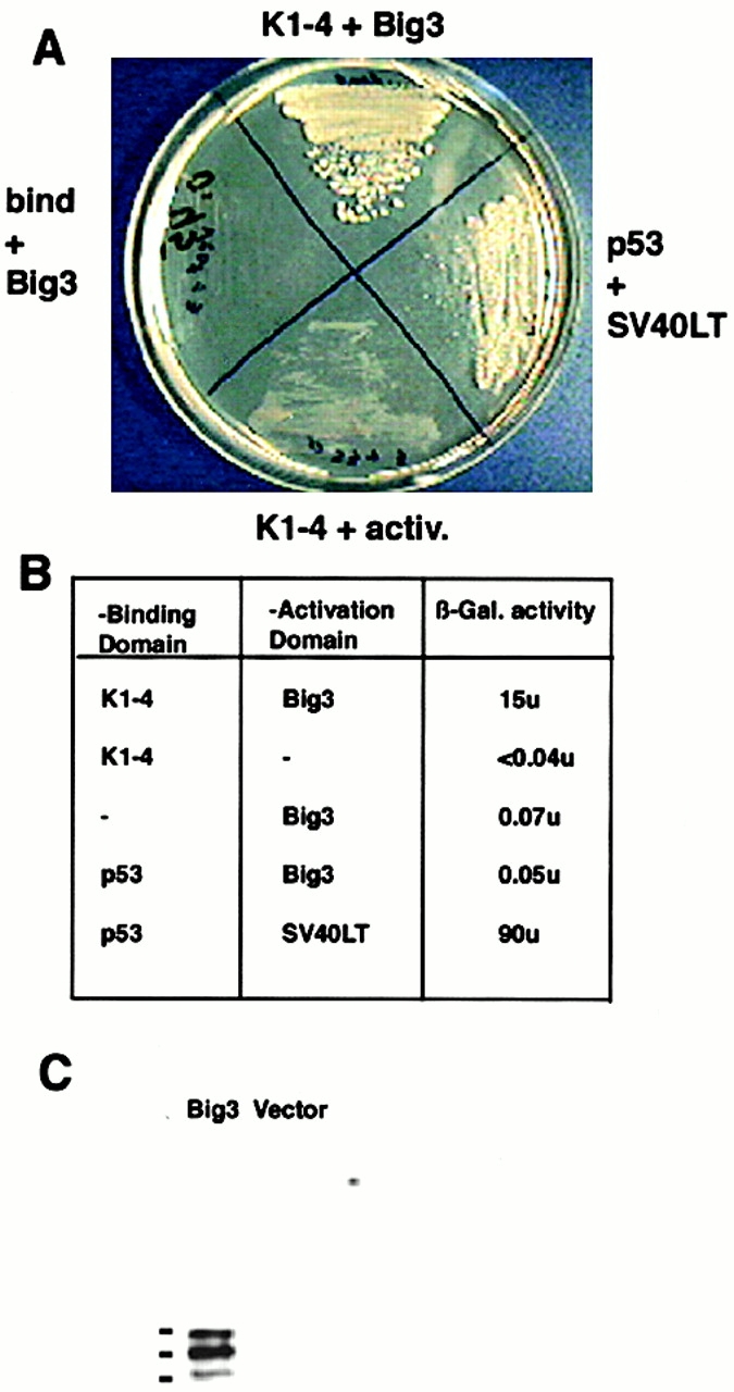
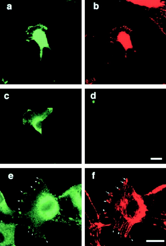
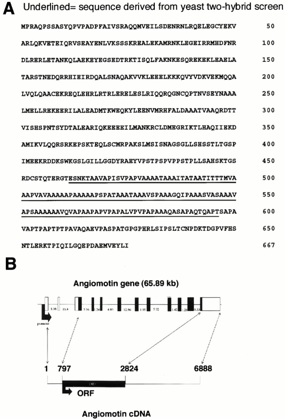
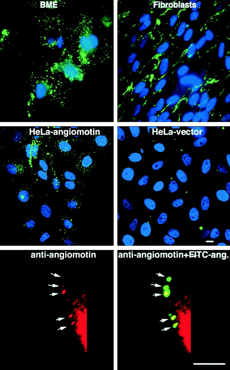
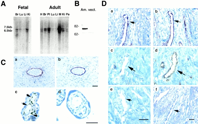
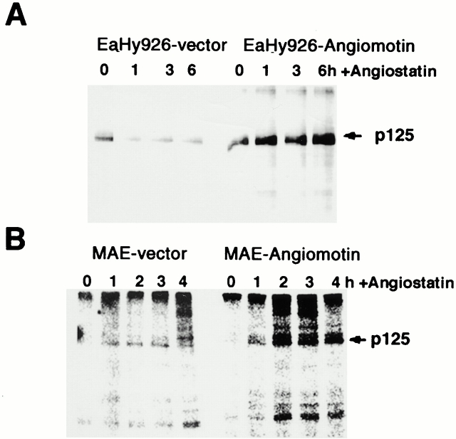
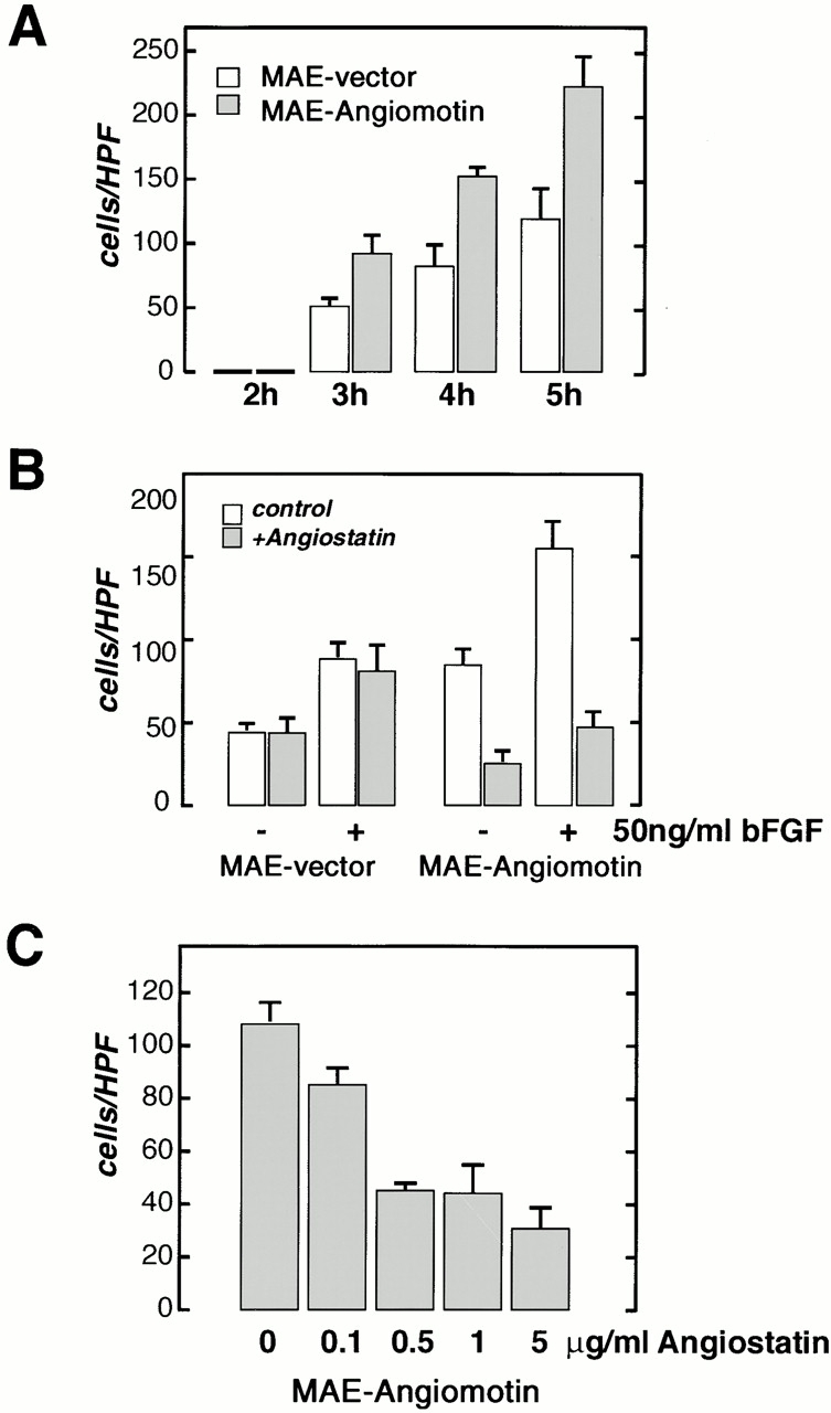
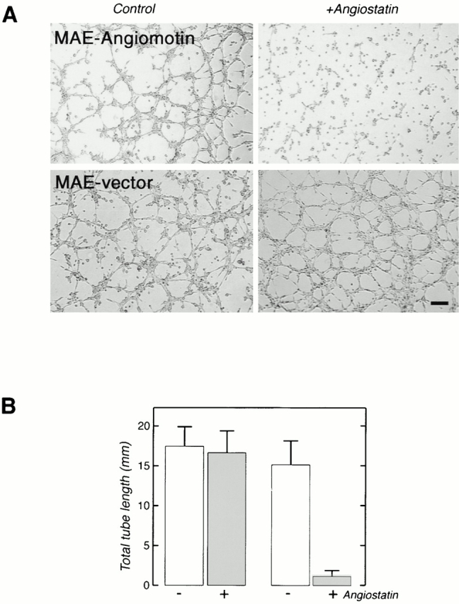
Angiostatin, a circulating inhibitor of angiogenesis, was identified by its ability to maintain dormancy of established metastases in vivo. In vitro, angiostatin inhibits endothelial cell migration, proliferation, and tube formation, and induces apoptosis in a cell type-specific manner. We have used a construct encoding the kringle domains 1--4 of angiostatin to screen a placenta yeast two-hybrid cDNA library for angiostatin-binding peptides. Here we report the identification of angiomotin, a novel protein that mediates angiostatin inhibition of migration and tube formation of endothelial cells. In vivo, angiomotin is expressed in the endothelial cells of capillaries as well as larger vessels of the human placenta. Upon expression of angiomotin in HeLa cells, angiomotin bound and internalized fluorescein-labeled angiostatin. Transfected angiomotin as well as endogenous angiomotin protein were localized to the leading edge of migrating endothelial cells. Expression of angiomotin in endothelial cells resulted in increased cell migration, suggesting a stimulatory role of angiomotin in cell motility. However, treatment with angiostatin inhibited migration and tube formation in angiomotin-expressing cells but not in control cells. These findings indicate that angiostatin inhibits cell migration by interfering with angiomotin activity in endothelial cells.


Figures

Figure 1
Figure 1
Yeast two-hybrid screening for angiostatin-binding proteins in a human placenta cDNA library. (A) Yeast transfected with the angiostatin-Gal4 binding domain and BIG3-Gal4 activation domain grow under growth-restricted conditions (−Leu, −His, −Trp) and in the presence of 50 mM 3-amino-1, 2,4-triazol. No growth was detected in yeast transfected with both the angiostatin-Gal4 binding domain and the Gal4 activation domain, or the Gal4 binding domain and the BIG3-Gal4 activation domain. The p53-Gal4 binding domain and SV40LT-Gal4 activation domain were used as positive controls. (B) The table shows the activity of the nonselectable β-gal marker in yeast cells transfected with angiostatin-Gal4 binding and the BIG3-Gal4 activation domain and controls. (C) Binding of the 421 amino acid BIG3 sequence derived from yeast two-hybrid screening was verified by coprecipitation of GST-BIG3 with angiostatin.
Figure 5
Figure 5
Subcellular localization of transfected and endogenous angiomotin. GFP-tagged angiomotin was expressed in NIH 3T3 cells using a retroviral expression system. Colocalization of GFP-angiomotin (a) with FAK (b) in the extending lamellipodium. (c) Immunofluorescence staining with a polyclonal antibody against angiomotin shows similar localization of endogenous angiomotin in a migrating HUVEC. (d) Control IgG from the same immunized animal. e and f shows the localization of angiomotin (e) to actin ruffles (f) and focal complexes in spreading HUVECs as visualized by phalloidin staining (arrows). Bars, 10 μm.
Figure 2
Figure 2
(A) The amino acid sequence of angiomotin. The amino acid sequence of the clone derived from the yeast two-hybrid screening is underlined. (B) Genomic organization of the angiomotin gene, size of cDNA, and the open reading frame (ORF). The Genbank accession number for the angiomotin cDNA genomic sequence data are available from GenBank/EMBL/DDBJ under accession nos. AF286598 and NT_011819.
Figure 3
Figure 3
Binding of FITC-labeled angiostatin to living cells in culture. Cells were incubated with 10 μg/ml FITC-labeled angiostatin at 4°C for 60 min. The cells were placed in 37°C incubator 15 min before washing in PBS and fixation in 3.7% formaldehyde. Angiostatin binds to and becomes internalized in bovine microcapillary endothelial (BME) cells resulting in a patchy staining of endosomes (top left). In contrast, angiostatin binds to the matrix of human fetal fibroblasts but is not internalized (top right). HeLa cells transfected with angiomotin bind and internalize angiostatin in a similar pattern to that of endothelial cells (middle left). HeLa-Ctrl cells are transfected with an empty vector and were negative for angiostatin binding (middle right). The bottom panel shows colocalization of FITC-angiostatin (ang.) with angiomotin during internalization after surface binding to HeLa cells transfected with angiomotin. Angiomotin was visualized by immunofluorescent staining using immunoaffinity-purified rabbit polyclonal antibodies against angiomotin as described in Materials and Methods. Bars, 10 μm.
Figure 4
Figure 4
Angiomotin is expressed in endothelial cells in vivo. (A) Northern blot analysis of angiomotin expression in fetal and adult human tissues. 2 μg of poly A+ RNA was analyzed for expression of angiomotin using a 1-kb probe from the 5′ region. Two detectable transcripts were detected in both fetal and adult tissues (multitissue blot from CLONTECH Laboratories, Inc.; H, heart; Br, brain; Pl placenta; Lu, lung; Li, liver; Ki, kidney; Pa, pancreas; M, marker). (B) Western blot analysis using immunoaffinity-purified rabbit polyclonal antibodies against angiomotin. The antibodies detected a band with an approximate molecular mass of 75 kD in angiomotin-transfected NIH 3T3 (Am.) cells but not in the vector (vect.) control. (C) Immunohistochemical localization of ABP-1 protein in human placenta. Paraffin-embedded placental tissue from term placenta was stained with immunoaffinity-purified angiomotin polyclonal antibodies. (C, a) Shows angiomotin protein in endothelium of larger vessels in a placental villus. (C, b) Immunostaining using a monoclonal antibody against CD34, an endothelial marker. (C, c) Detection of angiomotin protein in capillaries of placental microvilli. (C, d) IgG negative control. (D) Expression of angiomotin in a dermal Kaposis sarcoma. (D, a) Shows angiomotin staining of a blood vessel within the Kaposis lesion. (D, b) CD34 staining of a consecutive section of D, panel a. (D, c) Angiomotin-negative dermal blood vessel (arrow) in the normal tissue adjacent to the Kaposis lesion. (D, d) Same as D, panel c, but stained with CD34. D, panels e and f, are negative controls stained with nonimmune rabbit IgG as a negative control. Bars, 40 μm.
Figure 6
Figure 6
Angiostatin induces FAK activity in vitro in cells transfected with angiomotin. (A) Angiomotin-negative EaHy926 or (B) MAE cells were transfected with angiomotin or empty vector. Cells were stimulated with angiostatin at the indicated time points. FAK was immunoprecipitated and subjected to in vitro kinase analysis as described in Materials and Methods. Arrows indicate the localization of p125 FAK.
Figure 7
Figure 7
Migration of MAE cells transfected with angiomotin or with the vector alone was studied in the modified Boyden chamber assay. (A) Analysis of spontaneous migration in the absence of chemotactic factor at different time points after start of the experiment. A higher number of angiomotin-expressing cells are migrating through the filter at all time points. Vertical axis, cells migrated per high power field (HPF). (B) Effect of angiostatin on migration of MAE-angiomotin and MAE vector cells with or without stimulation with bFGF. Cells were pretreated for 1 h with angiostatin as indicated. (C) Dose response of angiostatin-mediated inhibition of migration of angiomotin-transfected MAE cells. All samples were performed in quadruplicates (error bars = SD).
Figure 8
Figure 8
(A) Angiostatin inhibits tube formation of angiomotin-transfected cells plated on matrigel. MAE-vector and MAE-angiomotin transfected cells were pretreated with 5 μg/ml angiostatin for 16 h before trypsinization and plating on matrigel. Images show tube formation 16 h after seeding on matrigel (Bar, 130 μm). (B) Total tube length formed in the presence or absence of angiostatin. The data represent the average from three independent experiments (error bars = SD).

Comment in

References

    1. Ahmed A., Li X.F., Dunk C., Whittle M.J., Rushton D.I., Rollason T. Colocalisation of vascular endothelial growth factor and its Flt-1 receptor in human placenta. Growth Factors. 1995;12:235–243. - PubMed
    1. Barendsz-Janson A.F., Griffioen A.W., Muller A.D., van Dam-Mieras M.C., Hillen H.F. In vitro tumor angiogenesis assaysplasminogen lysine binding site 1 inhibits in vitro tumor-induced angiogenesis. J. Vasc. Res. 1998;35:109–114. - PubMed
    1. Bastaki M., Nelli E.E, Dell'Era P., Rusnati M., Molinari-Tosatti M.P., Parolini S., Auerbach R., Ruco L.P., Possati L., Presta M. Basic fibroblast growth factor-induced angiogenic phenotype in mouse endothelium. A study of aortic and microvascular endothelial cell lines. Arterioscler. Thromb. Vasc. Biol. 1997;17:454–464. - PubMed
    1. Bergers G., Javaherian K., Lo K.M., Folkman J., Hanahan D. Effects of angiogenesis inhibitors on multistage carcinogenesis in mice. Science. 1999;284:808–812. - PubMed
    1. Bouck N., Stellmach V., Hsu S.C. How tumors become angiogenic. Adv. Cancer Res. 1996;69:135–174. - PubMed

Publication types

MeSH terms

Associated data