. 2001 Mar 19;193(6):671-8.
doi: 10.1084/jem.193.6.671.

A dendritic cell-specific intercellular adhesion molecule 3-grabbing nonintegrin (DC-SIGN)-related protein is highly expressed on human liver sinusoidal endothelial cells and promotes HIV-1 infection

Affiliations

A dendritic cell-specific intercellular adhesion molecule 3-grabbing nonintegrin (DC-SIGN)-related protein is highly expressed on human liver sinusoidal endothelial cells and promotes HIV-1 infection

A A Bashirova et al. J Exp Med. .

Abstract

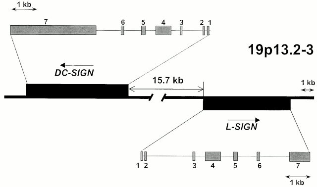
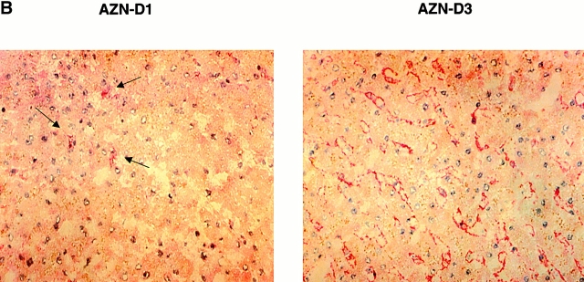
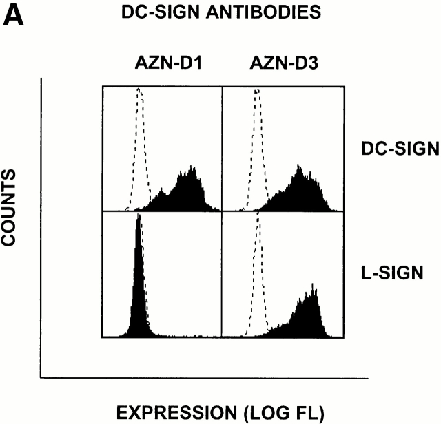
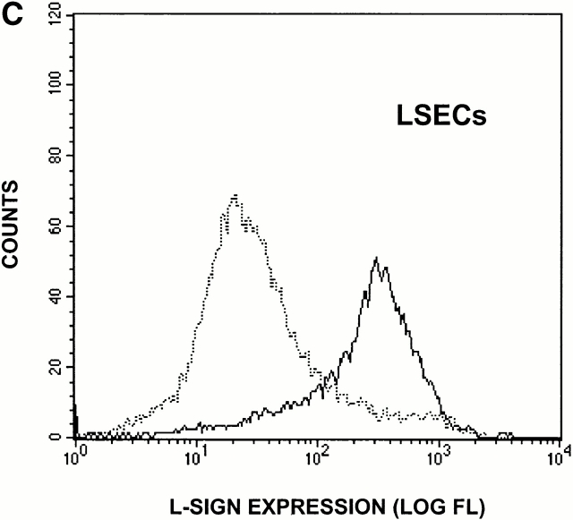
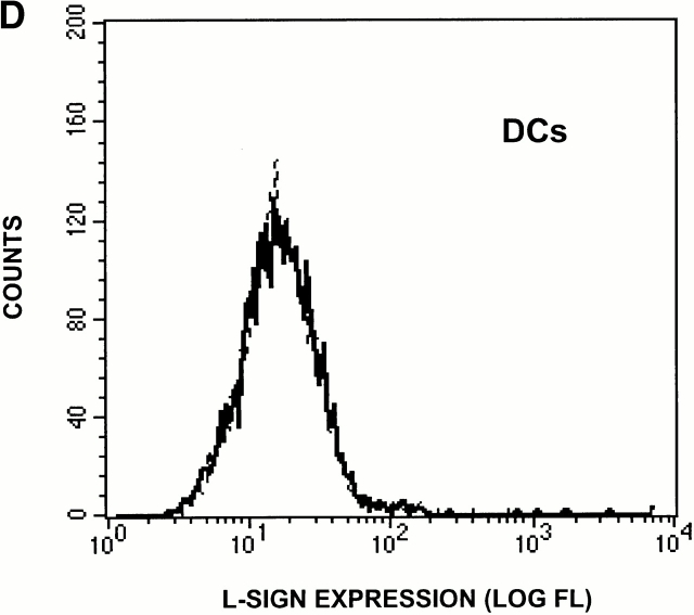
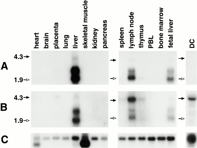
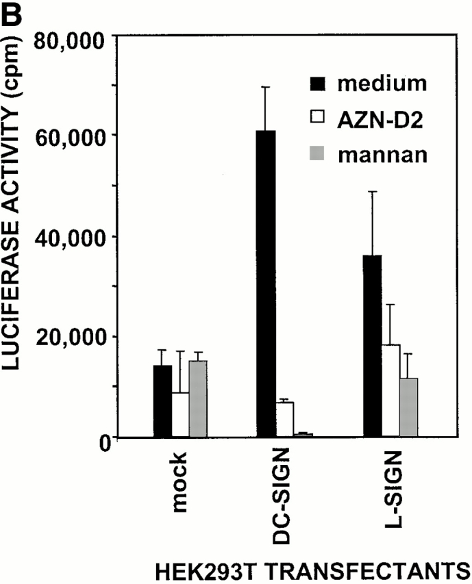
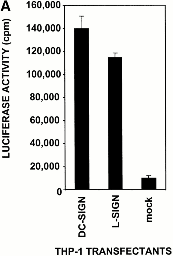
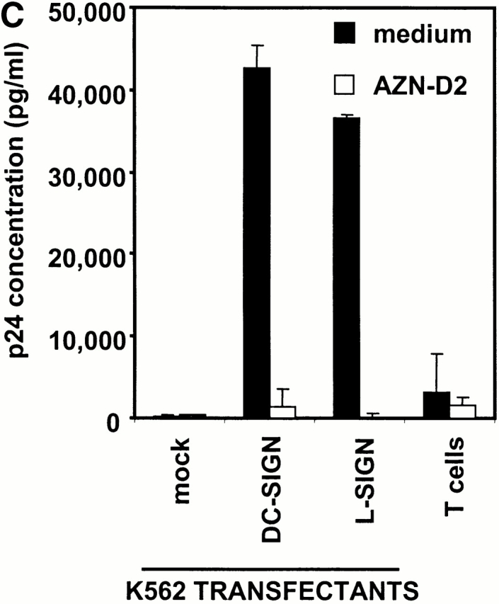
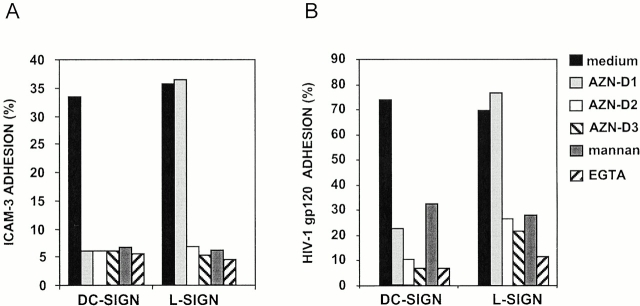
The discovery of dendritic cell (DC)-specific intercellular adhesion molecule (ICAM)-3-grabbing nonintegrin (DC-SIGN) as a DC-specific ICAM-3 binding receptor that enhances HIV-1 infection of T cells in trans has indicated a potentially important role for adhesion molecules in AIDS pathogenesis. A related molecule called DC-SIGNR exhibits 77% amino acid sequence identity with DC-SIGN. The DC-SIGN and DC-SIGNR genes map within a 30-kb region on chromosome 19p13.2-3. Their strong homology and close physical location indicate a recent duplication of the original gene. Messenger RNA and protein expression patterns demonstrate that the DC-SIGN-related molecule is highly expressed on liver sinusoidal cells and in the lymph node but not on DCs, in contrast to DC-SIGN. Therefore, we suggest that a more appropriate name for the DC-SIGN-related molecule is L-SIGN, liver/lymph node-specific ICAM-3-grabbing nonintegrin. We show that in the liver, L-SIGN is expressed by sinusoidal endothelial cells. Functional studies indicate that L-SIGN behaves similarly to DC-SIGN in that it has a high affinity for ICAM-3, captures HIV-1 through gp120 binding, and enhances HIV-1 infection of T cells in trans. We propose that L-SIGN may play an important role in the interaction between liver sinusoidal endothelium and trafficking lymphocytes, as well as function in the pathogenesis of HIV-1.


Figures

Figure 1
Figure 1
Schematic representation of the DC-SIGN/L-SIGN genetic map. Physical distances and gene orientation are based on the sequence provided from BAC clone CTD-2102F19 (GenBank/EMBL/DDBJ accession no. AC008812).
Figure 3
Figure 3
L-SIGN is expressed on LSECs and not on monocyte-derived DCs. (A) The antibody AZN-D1 is DC-SIGN specific, whereas AZN-D3 cross-reacts with L-SIGN. Stable DC-SIGN and L-SIGN K562 transfectants were stained with either AZN-D1 or AZN-D3. (B) Immunohistochemical analysis of DC-SIGN and L-SIGN expression in the human liver. Serial sections were stained with either AZN-D1 (DC-SIGN specific) or with AZN-D3 (detects both DC-SIGN and L-SIGN). AZN-D1 stains infrequent cells that may be DCs (arrows), whereas AZN-D3 stains cells lining sinusoids. (C) Expression of L-SIGN in liver is restricted to LSECs. 1 d after isolation, primary human liver cells were incubated with fluorochrome-labeled OVA. L-SIGN expression was determined by indirect immunofluorescence using an L-SIGN–specific polyclonal antibody. Cells that have taken up OVA (LSECs) and those that did not take up OVA (hepatocytes and other resident hepatic cells) are represented by solid and broken lines, respectively, by gating on the respective cell populations. 2 × 10⁵ cells were analyzed. (D) L-SIGN is not expressed by monocyte-derived DCs. Immature DCs, cultured from monocytes in the presence of GM-CSF and IL-4, do not stain with anti–L-SIGN polyclonal antibody, as determined by FACScan™ analysis. Solid line indicates staining with anti–L-SIGN polyclonal serum, whereas broken line (hidden under solid lane) represents staining with rabbit preimmune serum.
Figure 3
Figure 3
L-SIGN is expressed on LSECs and not on monocyte-derived DCs. (A) The antibody AZN-D1 is DC-SIGN specific, whereas AZN-D3 cross-reacts with L-SIGN. Stable DC-SIGN and L-SIGN K562 transfectants were stained with either AZN-D1 or AZN-D3. (B) Immunohistochemical analysis of DC-SIGN and L-SIGN expression in the human liver. Serial sections were stained with either AZN-D1 (DC-SIGN specific) or with AZN-D3 (detects both DC-SIGN and L-SIGN). AZN-D1 stains infrequent cells that may be DCs (arrows), whereas AZN-D3 stains cells lining sinusoids. (C) Expression of L-SIGN in liver is restricted to LSECs. 1 d after isolation, primary human liver cells were incubated with fluorochrome-labeled OVA. L-SIGN expression was determined by indirect immunofluorescence using an L-SIGN–specific polyclonal antibody. Cells that have taken up OVA (LSECs) and those that did not take up OVA (hepatocytes and other resident hepatic cells) are represented by solid and broken lines, respectively, by gating on the respective cell populations. 2 × 10⁵ cells were analyzed. (D) L-SIGN is not expressed by monocyte-derived DCs. Immature DCs, cultured from monocytes in the presence of GM-CSF and IL-4, do not stain with anti–L-SIGN polyclonal antibody, as determined by FACScan™ analysis. Solid line indicates staining with anti–L-SIGN polyclonal serum, whereas broken line (hidden under solid lane) represents staining with rabbit preimmune serum.
Figure 2
Figure 2
Northern blot analysis of DC-SIGN and L-SIGN. Positions of the 4.3-kb (black arrows) and 1.9-kb (white arrows) sizes are marked on the left. (A) Hybridization with the L-SIGN–specific probe indicating expression of the gene in liver, lymph node, and weakly in thymus. (B) Hybridization with the probe recognizing both genes. 4.3-kb bands represent DC-SIGN mRNA. The light upper band (∼4.2 kb) evident in liver and lymph node using the L-SIGN–specific probe (Fig. 3 A) is distinct from DC-SIGN mRNA (4.3 kb) due to the specificity of the probe, intensity patterns, and slight differences in size. (C) Reprobing of the blots with the β-actin cDNA control probe.
Figure 5
Figure 5
L-SIGN captures and enhances infection of T cells with HIV-1 in trans. (A) L-SIGN captures HIV-1 and transmits it to target cells. Stable DC-SIGN– or L-SIGN–expressing THP-1 transfectants were preincubated with HIV-luc/JRFL pseudovirions to allow capture of the virus. Cells were washed and THP-1 transfectants were cocultured with Hut/CCR5 target cells. Cell lysates were obtained after 3 d and analyzed for luciferase activity. For each of the coculture conditions employed, mock infected controls were uniformly <100 cps in activity. Each data set represents the mean of four separate wells of infected cells. One representative experiment out of two is shown. (B) L-SIGN enhances infection of T cells by pseudotyped HIV-1. HEK293T cells were transiently transfected with cDNA encoding DC-SIGN, L-SIGN, or empty vector. Control cells were preincubated with 20 μg/ml AZN-D2 or 20 μg/ml mannan. Low amounts of pseudotyped HIV-1ADA were added together with activated T cells as described previously (reference 1). Infectivity was determined after 2 d by measuring luciferase activity. One representative experiment of two performed is shown. Each experiment was done in triplicate wells. (C) L-SIGN enhances infection of T cells by replication competent HIV-1. Stable K562 transfectants of both L-SIGN and DC-SIGN were incubated with low virus concentrations of replication-competent M-tropic strain HIV-1JR-CSF (TCID50 100/ml). To determine the specificity, cells were preincubated with AZN-D2 (20 μg/ml). After 2 h, activated T cells were added as described previously (reference 2). Culture supernatants were collected at day 14 after K562-T cell coculture and HIV-1 production was measured using ELISA to determine p24 antigen levels. In control experiments, the same amount of virus was added directly to T cells. One representative experiment out of three is shown. Each data set represents the mean of three separate wells of infected cells.
Figure 5
Figure 5
L-SIGN captures and enhances infection of T cells with HIV-1 in trans. (A) L-SIGN captures HIV-1 and transmits it to target cells. Stable DC-SIGN– or L-SIGN–expressing THP-1 transfectants were preincubated with HIV-luc/JRFL pseudovirions to allow capture of the virus. Cells were washed and THP-1 transfectants were cocultured with Hut/CCR5 target cells. Cell lysates were obtained after 3 d and analyzed for luciferase activity. For each of the coculture conditions employed, mock infected controls were uniformly <100 cps in activity. Each data set represents the mean of four separate wells of infected cells. One representative experiment out of two is shown. (B) L-SIGN enhances infection of T cells by pseudotyped HIV-1. HEK293T cells were transiently transfected with cDNA encoding DC-SIGN, L-SIGN, or empty vector. Control cells were preincubated with 20 μg/ml AZN-D2 or 20 μg/ml mannan. Low amounts of pseudotyped HIV-1ADA were added together with activated T cells as described previously (reference 1). Infectivity was determined after 2 d by measuring luciferase activity. One representative experiment of two performed is shown. Each experiment was done in triplicate wells. (C) L-SIGN enhances infection of T cells by replication competent HIV-1. Stable K562 transfectants of both L-SIGN and DC-SIGN were incubated with low virus concentrations of replication-competent M-tropic strain HIV-1JR-CSF (TCID50 100/ml). To determine the specificity, cells were preincubated with AZN-D2 (20 μg/ml). After 2 h, activated T cells were added as described previously (reference 2). Culture supernatants were collected at day 14 after K562-T cell coculture and HIV-1 production was measured using ELISA to determine p24 antigen levels. In control experiments, the same amount of virus was added directly to T cells. One representative experiment out of three is shown. Each data set represents the mean of three separate wells of infected cells.
Figure 4
Figure 4
L-SIGN binds ICAM-3 (A) and HIV-1 gp120 (B). Adhesion of ICAM-3 and gp120 to the K562–L-SIGN and K562–DC-SIGN cells was measured with the fluorescent bead adhesion assay (reference 11). The y-axis represents the percentage of cells binding ligand-coated fluorescent beads. The L-SIGN cross-reacting mAbs AZN-D2 (20 μg/ml) and AZN-D3 (20 μg/ml) inhibit the adhesion of ICAM-3 and gp120 to L-SIGN, in contrast to the DC-SIGN–specific mAb AZN-D1 (20 μg/ml). Adhesion of both ICAM-3 and gp120 to the K562 transfectants is also inhibited by either 20 μg/ml mannan or 5 mM EGTA. Adhesion of both ligands to mock transfectants was <5%. One representative experiment out of three is shown (SD < 5%).

References

    1. Geijtenbeek T.B., Kwon D.S., Torensma R., van Vliet S.J., van Duijnhoven G.C., Middel J., Cornelissen I.L., Nottet H.S., KewalRamani V.N., Littman D.R. DC-SIGN, a dendritic cell-specific HIV-1-binding protein that enhances trans-infection of T cells. Cell. 2000;100:587–597. - PubMed
    1. Geijtenbeek T.B., Torensma R., van Vliet S.J., van Duijnhoven G.C., Adema G.J., van Kooyk Y., Figdor C.G. Identification of DC-SIGN, a novel dendritic cell-specific ICAM-3 receptor that supports primary immune responses. Cell. 2000;100:575–585. - PubMed
    1. Curtis B.M., Scharnowske S., Watson A.J. Sequence and expression of a membrane-associated C-type lectin that exhibits CD4-independent binding of human immunodeficiency virus envelope glycoprotein gp120. Proc. Natl. Acad. Sci. USA. 1992;89:8356–8360. - PMC - PubMed
    1. Yokoyama-Kobayashi M., Yamaguchi T., Sekine S., Kato S. Selection of cDNAs encoding putative type II membrane proteins on the cell surface from a human full-length cDNA bank. Gene. 1999;228:161–167. - PubMed
    1. Soilleux E.J., Barten R., Trowsdale J. DC-SIGN, a related gene DC-SIGNR, and CD23 form a cluster on 19p13. J. Immunol. 2000;165:2937–2942. - PubMed

Publication types

MeSH terms

Associated data