. 2001 Mar 19;193(6):689-98.
doi: 10.1084/jem.193.6.689.

Evidence that gammadelta versus alphabeta T cell fate determination is initiated independently of T cell receptor signaling

Affiliations

Evidence that gammadelta versus alphabeta T cell fate determination is initiated independently of T cell receptor signaling

J Kang et al. J Exp Med. .

Abstract

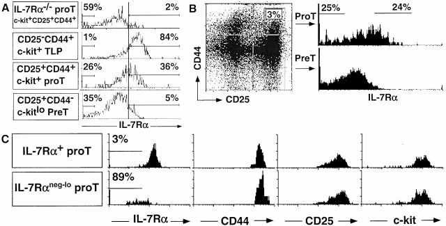
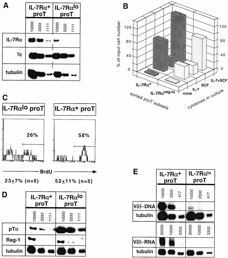
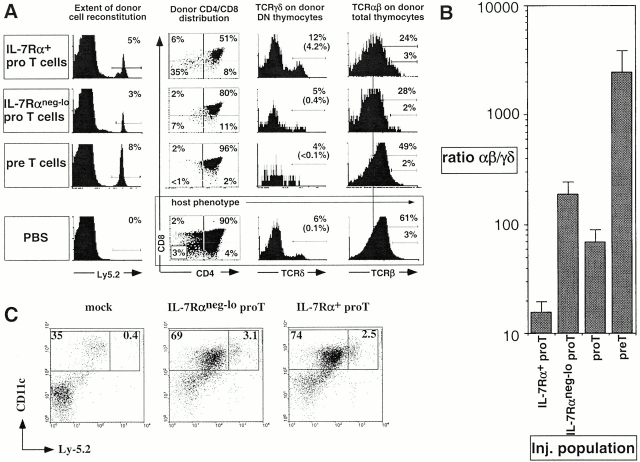
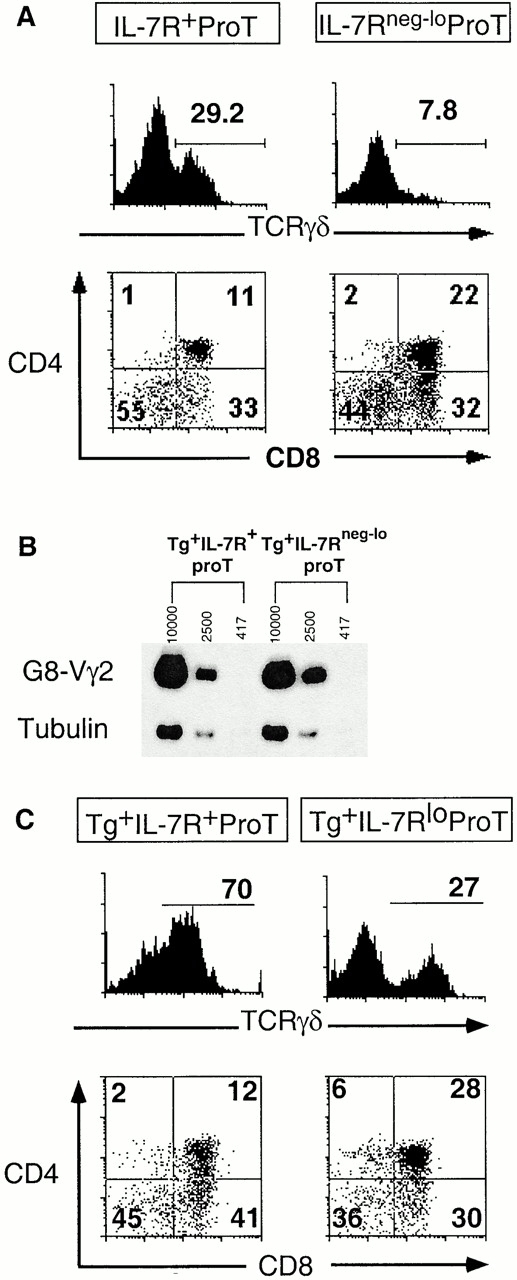
Two types of T cells, alphabeta and gammadelta, develop in vertebrates. How these two T cell lineages arise from a common thymic T progenitor is poorly understood. Differentiation of alphabeta lineage T cells requires the surrogate alpha chain (pTalpha), which associates with the T cell receptor (TCR) beta chain to form the pre-TCR. gammadelta lineage development does not appear to involve an obligatory surrogate chain, but instead requires productive rearrangement and expression of both TCR gamma and delta genes. It has been proposed that the quality of signals transmitted by the pre-TCR and gammadelta TCR are distinct and that these "instructive" signals determine the lineage fate of an uncommitted progenitor cell. Here we show that the thymic T progenitor cells (CD25(+)CD44(+)c-kit(+)CD3(-)CD4(-)CD8(-) thymocytes, termed pro-T cells) from young adult mice that have yet to express TCRs can be subdivided based on interleukin 7 receptor (IL-7R) expression. These subsets exhibit differential potential to develop into gammadelta versus alphabeta lineage (CD4+CD8+ cells) in the thymus. Upon intrathymic injection, IL-7R(neg-lo) pro-T cells generated a 13-fold higher ratio of alphabeta lineage to gammadelta lineage cells than did IL-7R(+) pro-T cells. Much of this difference was due to a fivefold greater potential of IL-7R(+) pro-T cells to develop into TCR-gammadelta T cells. Evidence indicates that this biased developmental potential is not a result of enhanced TCR-gamma gene rearrangement/expression in IL-7R(+) pro-T cells. These results indicate that the pro-T cells are heterogeneous in developmental potential before TCR gene rearrangement and suggest that in some precursor cells the initial lineage commitment is independent of TCR-mediated signals.


Figures

Figure 1
Figure 1
Subdivision of pro-T cells based on IL-7Rα expression. (A) CD4CD8 thymocytes from B6 and B6 IL-7Rα−/− mice were stained with mAbs for CD25, CD44, IL-7Rα (B12-1), and c-kit. IL-7Rα chain levels on indicated precursor subsets are shown. The level of positive staining for IL-7Rα chain was determined by comparing to the fluorescence signal from IL-7Rα−/− thymocytes (top). An irrelevant, isotype-matched (to B12-1) control mAb did not stain T precursor subsets and CD25+ cells were not stained by mAb specific for CD3ε chain (data not shown). (B) Sorting gates for IL-7R+ and IL-7Rneg-lo CD44+CD25+ TN pro-T cells are shown. IL-7Rα staining of CD25+CD44 TN pre-T cells is presented for comparison. (C) Postsort analysis of similar numbers of purified populations with the indicated mAbs. Sorted cells were additionally stained with anti–c-kit–APC and analyzed on an ELITE flow cytometer. Many of the IL7Rαneg-lo cells in A and C accumulate in the lowest fluorescence intensity channel, characteristic of Beckman Coulter flow cytometers, especially when using four-color compensation settings.
Figure 2
Figure 2
IL-7Rα+ and IL-7Rαneg-lo pro-T subsets exhibit distinct properties. (A) A representative radioactive RT-PCR analysis for IL-7Rα, γc, and tubulin transcripts in the sorted pro-T subsets of similar purity. Numbers indicate approximate cell equivalents used for IL-7Rα and γc-specific PCR. For tubulin PCR, the starting concentration was 3,333 cell equivalents, which was subject to two serial threefold dilutions. The IL-7Rα and γc autoradiographs were exposed for 3 d or overnight, respectively. No PCR products were detected when the RT step was omitted. (B) Proportion of input sorted cells surviving culturing the pro-T subsets for 4 d in the presence of IL-7 and/or SCF. (C) BrdU incorporation after a 3-h pulse with BrdU in vivo. Treated DN thymocytes were stained with mAbs for CD25-613, CD44-Cy5, IL-7Rα-biotin/strepavidin-PE, and BrdU-FITC. The levels of BrdU staining on gated CD25+CD44+IL-7Rα+ and CD25+CD44+IL-7Rαneg-lo TN thymocytes are presented. (D) RT-PCR analysis for pTα and Rag-1 transcripts. Results from one of four independent sorting experiments are shown for pTα expression analysis; two experiments showed a marginal difference (two- to threefold), whereas two others showed a larger (more than ninefold) difference. (E) Levels of Vδ5-Jδ1 rearrangements in genomic DNA (top) and Vδ5-Jδ1 transcripts in total RNA (bottom) from sorted pro-T cell subsets, determined by semiquantitative PCR or RT-PCR, respectively. Genomic DNA PCR was for 35 cycles; RT-PCR entailed 38 cycles for Vδ5-Jδ1 and 28 cycles for tubulin. No PCR products were detected when the RT step was omitted. The autoradiographs shown for Vδ5-Jδ1 and tubulin transcripts were exposed for 5 d and 4 h, respectively.
Figure 3
Figure 3
Intrathymically injected pro-T cell subsets displayed distinct γδ versus αβ lineage developmental potential. (A) Representative profiles of thymocytes generated from donor (Ly5.2+) precursor cells. CD4/CD8 profiles of donor-type thymocytes, γδ TCR expression on gated donor-type CD4CD8 thymocytes, and αβ TCR expression on donor thymocytes are illustrated. Host thymocyte profiles are presented for a mouse injected with saline alone. Percentages in brackets represent the percentage of γδ TCR+ cells of all donor thymocytes. In the profiles compared, IL-7Rα1 and IL-7Rαneg-lo pro-T cells generated similar numbers of donor-derived thymocytes. (B) The ratio of αβ to γδ thymocytes generated from the respective sorted populations 8–11 d after injection. The bars represent averages of αβ/γδ thymocyte ratio of individual mice (see Table ). Total donor thymocyte numbers were higher in the thymi injected with IL-7Rneg-lo pro-T cells, but the broad range (0.2–10.6%) of donor cell reconstitution level in each experimental group makes the interpretation of this difference ambiguous. (C) Representative profiles of five independent experiments show similar proportions of DCs generated from the pro-T subsets (average proportions of donor DCs in CD11c+ population ± SEM: IL-7Rαneg-lo, 10.4 ± 2.6%; IL-7Rα+, 12.2 ± 2.4%). Thymic DCs were purified from mice 9 d after intrathymic injection of pro-T cells or no cells (mock), as indicated. The cells were analyzed for CD11c expression, which identifies all DCs, and for Ly-5.2 expression, which distinguishes DCs of donor (Ly-5.2+) versus host (Ly-5.2) origin. Purified DCs (all MHC class II+) from mice with 4% and 1.5–1.9% donor cell reconstitution from injected IL-7Rαneg-lo and IL-7Rα+ pro-T cells, respectively, are shown. For comparison, cells from a mouse that underwent the same surgical procedure but was not injected are presented (mock).
Figure 4
Figure 4
The sorted pro-T subsets display distinct γδ/αβ lineage developmental potential in FTOC. (A) Thymocytes from FTOCs reconstituted with sorted pro-T cell subsets were analyzed for γδ TCR and CD4/CD8 expression after 11 d of culture. Representative profiles are from three thymic lobes that were pooled. Many of the cells are accumulating on the axes of the profiles as a result of four-color compensation settings. (B) Similar transgene expression in sorted pro-T cell subsets from low 1 2 transgene copy line as determined by semiquantitative RT-PCR using transgene-specific primers. Numbers indicate approximate cell equivalents. Two additional experiments yielded comparable results. No PCR products were detected when the RT step was omitted. (C) Thymocytes from FTOCs reconstituted with sorted pro-T cell subsets from high copy G8 transgenic mice were analyzed for γδ TCR and CD4/CD8 expression after 10 d of culture. The levels of IL-7Rα expression on the pro-T progenitor populations in the transgenic mice were not notably different from those in nontransgenic littermates. Three thymic lobes of each type were pooled for analysis.

References

    1. Fehling H., Krotkova A., Saint-Ruf C., von Boehmer H. Crucial role of the pre-T cell receptor α gene in the development of αβ but not γδ T cells. Nature. 1995;375:795–798. - PubMed
    1. Dudley E.C., Petrie H.T., Shah L.M., Owen M.J., Hayday A.C. T cell receptor β chain gene rearrangement and selection during thymocyte development in adult mice. Immunity. 1994;1:83–93. - PubMed
    1. Penit C., Lucas B., Vasseur F. Cell expansion and growth arrest phases during the transition from precursor (CD4−8−) to immature (CD4+8+) thymocytes in normal and genetically modified mice. J. Immunol. 1995;154:5103–5113. - PubMed
    1. Kang J., Coles M., Cado D., Raulet D.H. The developmental fate of T cells is critically influenced by TCR γδ expression. Immunity. 1998;8:427–438. - PubMed
    1. Allison J.P., Lanier L.L. The T-cell antigen receptor γ generearrangement and cell lineages. Immunol. Today. 1987;8:293–296. - PubMed

Publication types

Substances