. 2001 Feb;2(2):102-7.
doi: 10.1093/embo-reports/kve018.

Molecular and cytological analysis of a 5.5 Mb minichromosome

Affiliations

Molecular and cytological analysis of a 5.5 Mb minichromosome

C Auriche et al. EMBO Rep. 2001 Feb.

Abstract

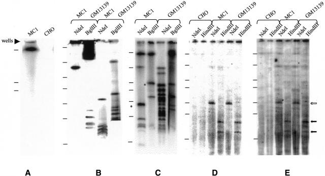
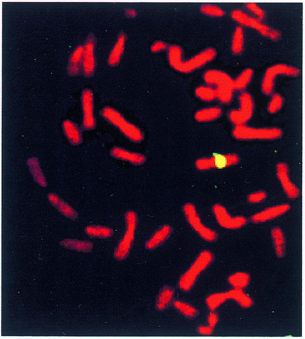
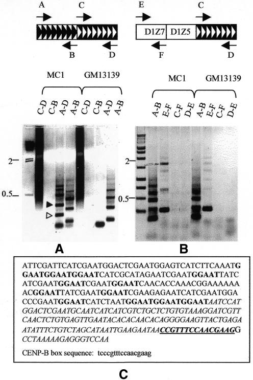
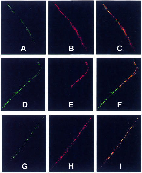
Mammalian artificial chromosomes (MACs) provide a new tool for the improvement of our knowledge of chromosome structure and function. Moreover, they constitute an alternative and potentially powerful tool for gene delivery both in cultured cells and for the production of transgenic animals. In the present work we describe the molecular structure of MC1, a human minichromosome derived from chromosome 1. By means of restriction and hybridization analysis, satellite-PCR, in situ hybridization on highly extended chromatin fibres, and indirect immunofluorescence, we have established that: (i) MC1 has a size of 5.5 Mb; (ii) it consists of 1.1 Mb alphoid, 3.5 Mb Sat2 DNA, and telomeric and subtelomeric sequences at both ends; (iii) it contains an unusual region of interspersed Sat2 and alphoid DNAs at the junction of the alphoid and the Sat2 blocks; and (iv) the two alphoid blocks and the Sat2-alphoid region bind centromeric proteins suggesting that they participate in the formation of a functional kinetochore.


Figures

None
Fig. 1. Long range mapping of MC1 and human chromosome 1. (A) undigested MC1 and CHO plugs hybridized to the Sat2 probe. (B–E) MC1 and GM13139 plugs digested with the indicated enzymes hybridized with: (B) chromosome 1 alphoid probe, (C) Sat2 DNA, (D) subtelomeric probe and (E) telomeric probe. The asterisk indicates double bands which were resolved under different run conditions. S. pombe chromosome markers in (A) were: 5.7, 4.6 and 3.5 Mb. Markers in (B–E) were: 2.2 Mb, 1.12 Mb, 750 kb, 610 kb, 450 kb, 225 kb and 48 kb. At the right side of the figure the filled and open arrows indicate HindIII and NdeI fragments, respectively.
None
Fig. 2. Reverse painting of GM13139 chromosomes with the biotin-labelled MC1 probe revealed with avidin-FITC. The chromosomes were counterstained with propidium iodide.
None
Fig. 3. Satellite-PCR mapping and sequencing. Gel electrophoresis of MC1 and GM13139 PCR obtained with the primers indicated at the top of each panel. Amplified region in (A) and (B): (A) alphoid/Sat2; (B) D1Z7-Sat2. (C) Sequence of the fragment marked with a filled triangle in (A); the GGAAT repeat, characteristic of Sat2 DNA is shown in bold; the region homologous to the alphoid DNA is italicized; the CENP-B box is underlined, and the consensus sequence is reported below in lower case. The first slot in (A) and (B) contains λ-HindIII size markers and a 1 kb ladder, respectively. The size markers indicated are in kb. Open and filled triangles in (A) indicate the two sequenced fragments. Symbols: black chevrons, alphoid block; white chevrons, Sat2 block; both oriented as in GenBank.
None
Fig. 4. Dual colour fibre-FISH of MC1. Extended chromatin fibres were simultaneously hybridized with: (A) pAL1–green/pZ5.1–red; (B) pZ5.1–red/Sat2–green; (C) pAL1–red/Sat2–green. The insert shows DAPI counterstained fibres, the white arrow points to the hybridized fibre. Digoxigenin-labelled pAL1 and Sat2 probes were revealed by anti-digoxigenin-FITC, biotin-labelled pZ5.1 and pAL1 were revealed by avidin-Cy3.
None
Fig. 5. Immunofluorescence/FISH analysis of MC1. Centromeric proteins were detected on extended chromatin fibres with (AF) CREST and (GI) anti-CENP-F, and subsequently the fibres were hybridized in situ to alphoid (A-C) and Sat2 (D-F) probes. CREST immunofluorescence (red) partially overlaps the Sat2 block (green), whereas it completely overlaps the alphoid region (green). CENP-F immunofluorescence (red) extends over the entire alphoid fibre (green). Merged images are shown in (C), (F) and (I). CREST and CENP-F were revealed with Cy3 and Texas-red, respectively; biotin-labelled Sat2 and alphoid probes with avidin-FITC.

Similar articles

Cited by

References

    1. Aleksandrov I.A., Akopian, T.A., Vinnik, E.A., Mitkevich, S.P. and Kiselev, L.L. (1988) Primary structure of chromosome-specific human alpha-satellite DNA. Dokl. Akad. Nauk SSSR, 300, 1235–1239. - PubMed
    1. Ascenzioni F., Della Valle, G., Guerrini, A.M., Pisani, G., Biondi, O. and Donini, P. (1990) A human DNA telomeric fragment containing yeast ARS and mitotic stabilizing sequences. Res. Microbiol., 141, 1117–1129. - PubMed
    1. Au H.C., Mascarello, J.T. and Scheffler, I.E. (1999) Targeted integration of a dominant neoR marker into a 2- to 3-Mb Human minichromosome and transfer between cells. Cytogenet. Cell Genet., 86, 194–203. - PubMed
    1. Bertoni L., Attolini, C., Faravelli, M., Simi, S. and Giulotto, E. (1996) Intrachromosomal telomere-like DNA sequences in Chinese hamster. Mamm. Genome, 7, 853–855. - PubMed
    1. Carine K., Solus, J., Waltzer, E., Manch-Citron, J., Hamkalo, B.A. and Scheffler, I.E. (1986) Chinese hamster cells with a minichromosome containing the centromeric region of human chromosome 1. Somat. Cell Mol. Genet., 12, 479–491. - PubMed

Publication types

MeSH terms

Associated data