. 2001 Apr;21(7):2281-91.
doi: 10.1128/MCB.21.7.2281-2291.2001.

Strong functional interactions of TFIIH with XPC and XPG in human DNA nucleotide excision repair, without a preassembled repairosome

Affiliations

Strong functional interactions of TFIIH with XPC and XPG in human DNA nucleotide excision repair, without a preassembled repairosome

S J Araújo et al. Mol Cell Biol. 2001 Apr.

Abstract

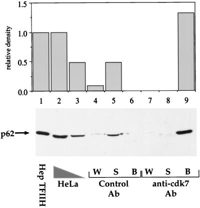
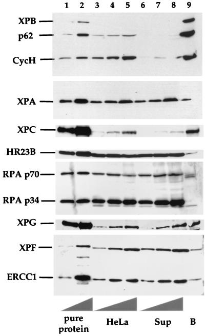
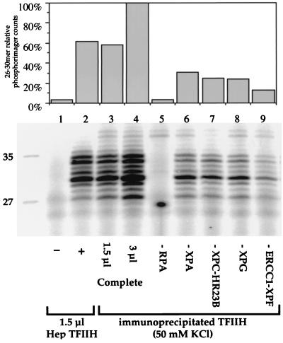
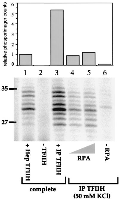
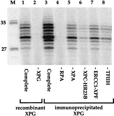
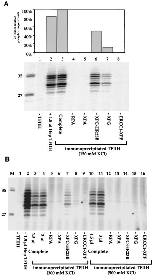
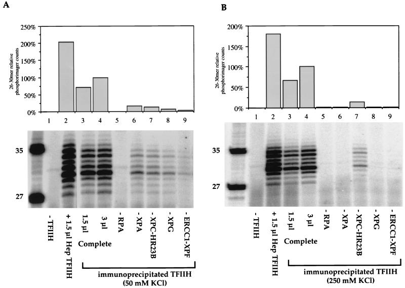
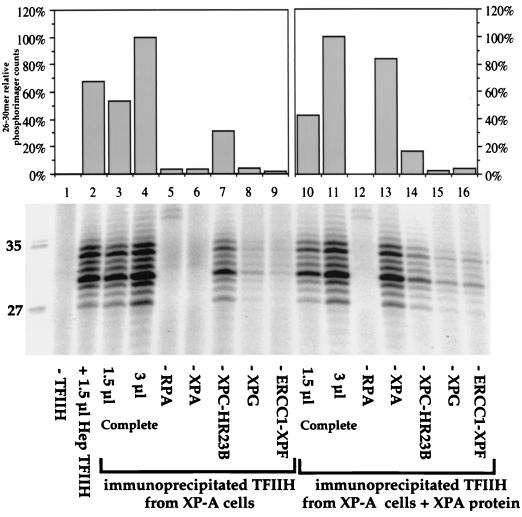
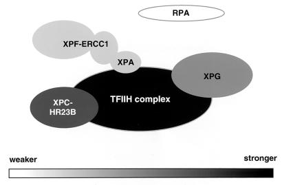
In mammalian cells, the core factors involved in the damage recognition and incision steps of DNA nucleotide excision repair are XPA, TFIIH complex, XPC-HR23B, replication protein A (RPA), XPG, and ERCC1-XPF. Many interactions between these components have been detected, using different physical methods, in human cells and for the homologous factors in Saccharomyces cerevisiae. Several human nucleotide excision repair (NER) complexes, including a high-molecular-mass repairosome complex, have been proposed. However, there have been no measurements of activity of any mammalian NER protein complex isolated under native conditions. In order to assess relative strengths of interactions between NER factors, we captured TFIIH from cell extracts with an anti-cdk7 antibody, retaining TFIIH in active form attached to magnetic beads. Coimmunoprecipitation of other NER proteins was then monitored functionally in a reconstituted repair system with purified proteins. We found that all detectable TFIIH in gently prepared human cell extracts was present in the intact nine-subunit form. There was no evidence for a repair complex that contained all of the NER components. At low ionic strength TFIIH could associate with functional amounts of each NER factor except RPA. At physiological ionic strength, TFIIH associated with significant amounts of XPC-HR23B and XPG but not other repair factors. The strongest interaction was between TFIIH and XPC-HR23B, indicating a coupled role of these proteins in early steps of repair. A panel of antibodies was used to estimate that there are on the order of 10(5) molecules of each core NER factor per HeLa cell.


Figures

FIG. 1
FIG. 1
Quantitative immunoprecipitation of TFIIH from HeLa cell extracts with a cdk7 antibody. TFIIH was immunoprecipitated from a HeLa whole-cell extract with a cdk7 antibody and detected using monoclonal antibody 3C9 against the p62 subunit. Lane 1 contains 5 μl of Hep TFIIH (fraction IV from HeLa cells), and lanes 2 and 3 contain 100 and 50 μg of HeLa whole-cell extract protein, respectively. HeLa cell extract protein (400 μg in 20 μl) was added to 40 μl of antibody (Ab) beads. Lanes 4 to 9 contain 20 μl of the supernatant (S) and first-wash (W) fractions and 10 μl of the bead (B) fraction. The control antibody was mouse whole IgG.
FIG. 2
FIG. 2
Coimmunoprecipitation of TFIIH and other NER factors. TFIIH was immunoprecipitated from a HeLa whole-cell extract, and TFIIH bound fractions were analyzed by immunoblotting. Detection was performed by using the indicated antibodies against proteins involved in the first steps of NER. Lanes 1 and 2 contain pure proteins (from top to bottom) as follows: 2.5 and 5 μl of Hep TFIIH, 22.5 and 45 ng of XPA, 75 and 150 ng of XPC-HR23B, 125 and 250 ng of RPA, 125 and 250 ng of XPG, and 50 and 100 ng of ERCC1-XPF. Lanes 3 to 5 contain 17.5, 35, and 70 μg of HeLa cell extract protein, respectively; lanes 6 to 8 contain 17.5, 35, and 70 μg of the supernatant protein from the same extract after immunoprecipitation (Sup); and lane 9 contains 20 μl of antibody beads (B) with the immunoprecipitate.
FIG. 3
FIG. 3
Functional interactions of TFIIH with other NER factors in HeLa cell extracts. cdk7 antibody beads containing TFIIH and associated proteins immunoprecipitated from HeLa cells were added to dual-incision assays reconstituted with purified factors. Protein-protein interactions were tested by omission of individual repair factors as indicated. Lanes 1 to 4 contain XPA, RPA, XPC-HR23B, XPG, and ERCC1-XPF, with no TFIIH (lane 1), 1.5 μl of Hep TFIIH (lane 2), or TFIIH beads (lanes 3 and 4). Excised fragments were detected by a direct end-labeling procedure which extends the 24 to 32-nucleotide products by 4 nucleotides. The quantification shown at the top was normalized to lane 4, containing 3 μl of TFIIH beads. TFIIH interactions in lanes 3 to 9 were measured on beads washed with buffer containing 50 mM KCl. Lanes 5 to 9 contain 3 μl of beads.
FIG. 4
FIG. 4
RPA restores NER activity to HeLa immunoprecipitates (IP). The results of dual-incision assays are presented as in Fig. 3. Lanes 1 to 3 contain XPA, RPA, XPC-HR23B, XPG, and ERCC1-XPF, with 1.5 μl of Hep TFIIH (lane 1) or no TFIIH (lane 2). Magnetic beads (10 μl) containing TFIIH and associated factors were washed with buffer containing 50 mM KCl and used in reconstituted dual-incision assays either with all the other repair factors added (lane 3), with only RPA added (lanes 4 to 5), or with no additions (lane 6). Lanes 4 and 5 contain 50 ng (0.45 pmol) and 125 ng (1.1 pmol) of RPA, respectively.
FIG. 5
FIG. 5
Functional interactions between XPG and other NER factors. XPG was immunoprecipitated from HeLa cell extracts, and the 8H7 antibody beads containing XPG and associated proteins were added to reconstituted dual-incision assays. Functional interactions were tested by omission of individual repair factors as indicated. Lanes marked “Complete” contain all the repair factors. Lane 1 contains 45 ng of purified recombinant XPG protein; lanes 3 to 8 contain 3 μl of XPG magnetic beads; and lane M contains molecular size markers, with lengths (in bases) noted at left.
FIG. 6
FIG. 6
Sensitivity of TFIIH interactions to ionic strength. cdk7 antibody beads containing TFIIH and associated proteins immunoprecipitated from HeLa cells were added to dual-incision assays reconstituted with purified factors. Protein-protein interactions were tested by omission of individual repair factors as indicated. (A) TFIIH interactions on beads washed with buffer containing 150 mM KCl. Lanes 1 to 3 contain XPA, RPA, XPC-HR23B, XPG, and ERCC1-XPF, with no TFIIH (lane 1), 1.5 μl of Hep TFIIH (lane 2), or TFIIH beads (lane 3). The quantification shown at the top was normalized to lane 3, containing 3 μl of TFIIH beads. Lanes 4 to 8 contain 3 μl of beads. (B) TFIIH interactions on beads washed with buffer containing 300 mM KCl (lanes 3 to 9) or 500 mM KCl (lanes 10 to 16). Lanes 1 to 4, 10, and 11 contain XPA, RPA, XPC-HR23B, XPG, and ERCC1-XPF, with no TFIIH (lane 1), 1.5 μl of Hep TFIIH (lane 2), 1.5 μl of TFIIH beads (lanes 3 and 10), or 3 μl of TFIIH beads (lanes 4 and 11). Lanes 5 to 16 contain 3 μl of beads. Lane M contains molecular size markers, with lengths (in bases) noted at left.
FIG. 7
FIG. 7
Functional interactions of TFIIH with other NER factors in extracts from normal lymphoblastoid cells. cdk7 magnetic beads containing TFIIH and associated proteins immunoprecipitated from 705ori cells were added to dual-incision assay mixtures reconstituted with purified factors. Protein-protein interactions were tested by omission of individual repair factors as indicated. Lanes marked “Complete” contain all the factors. (A) TFIIH interactions on beads washed with buffer containing 50 mM KCl. (B) TFIIH interactions on beads washed with buffer containing 250 mM KCl. In each case, lane 2 contains 1.5 μl of Hep TFIIH; lanes 3 and 4 contain 1.5 and 3 μl of cdk7 magnetic beads, respectively; and lanes 5 to 9 contain 3 μl of beads.
FIG. 8
FIG. 8
Functional interactions of TFIIH with other NER factors in extracts from XP-A cells. cdk7 magnetic beads containing TFIIH and associated proteins immunoprecipitated from GM2345 XP-A cells were washed with buffer containing 50 mM KCl and added to dual-incision assays reconstituted with purified factors. Protein-protein interactions were tested by omission of individual repair factors as indicated. Lanes marked “Complete” contain all the repair factors. Lanes 3 to 9 contain reaction mixtures with TFIIH immunoprecipitated from the XP-A cell extracts; lanes 10 to 16 contain reaction mixtures with TFIIH immunoprecipitated from the XP-A extracts after addition of 1 ng of purified XPA per μg of cell extract protein and incubation for 30 min at 30°C. Lane 2 contains 1.5 μl of Hep TFIIH, lanes 3 and 10 contain 1.5 μl of TFIIH antibody beads, and lanes 4 to 9 and 11 to 16 contain 3 μl of beads. Quantification was performed on a phosphorimager relatively to the lane that contains 3 μl of TFIIH beads (lane 4 for the first set of values and lane 11 for the second set).
FIG. 9
FIG. 9
Protein-protein interactions between human NER factors. Presented is a summary of the most stable interactions between core NER factors in human cell extracts; darker tones represent stronger interactions with TFIIH, and lighter tones represent weaker interactions with TFIIH, as detected by functional assays in the present study.

References

    1. Adamczewski J P, Rossignol M, Tassan J P, Nigg E A, Moncollin V, Egly J M. MAT1, cdk7 and cyclin H form a kinase complex which is UV light-sensitive upon association with TFIIH. EMBO J. 1996;15:1877–1884. - PMC - PubMed
    1. Araújo S J, Tirode F, Coin F, Pospiech H, Syväoja J E, Stucki M, Hübscher U, Egly J-M, Wood R D. Nucleotide excision repair of DNA with recombinant human proteins: definition of the minimal set of factors, active forms of TFIIH and modulation by CAK. Genes Dev. 2000;14:349–359. - PMC - PubMed
    1. Araújo S J, Wood R D. Protein complexes in nucleotide excision repair. Mutat Res. 1999;435:23–33. - PubMed
    1. Bardwell A J, Bardwell L, Iyer N, Svejstrup J Q, Feaver W J, Kornberg R D, Friedberg E C. Yeast nucleotide excision repair proteins Rad2 and Rad4 interact with RNA polymerase II basal transcription factor b (TFIIH) Mol Cell Biol. 1994;14:3569–3576. - PMC - PubMed
    1. Batty D P, Otrin V R, Levine A S, Wood R D. Stable binding of human XPC-hHR23B complex to irradiated DNA confers strong discrimination for damaged sites. J Mol Biol. 2000;300:275–290. - PubMed

Substances