. 2001 Apr;21(7):2373-83.
doi: 10.1128/MCB.21.7.2373-2383.2001.

p38 mitogen-activated protein kinase-dependent activation of protein phosphatases 1 and 2A inhibits MEK1 and MEK2 activity and collagenase 1 (MMP-1) gene expression

Affiliations

p38 mitogen-activated protein kinase-dependent activation of protein phosphatases 1 and 2A inhibits MEK1 and MEK2 activity and collagenase 1 (MMP-1) gene expression

J Westermarck et al. Mol Cell Biol. 2001 Apr.

Abstract

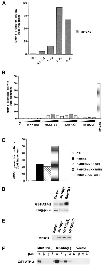
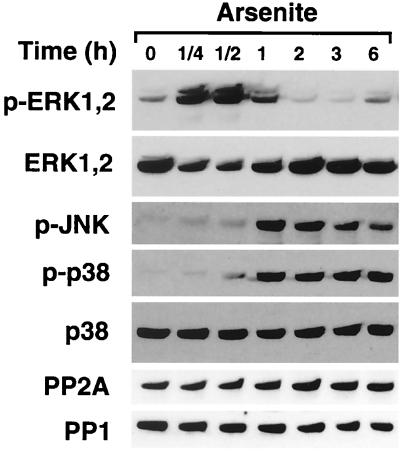
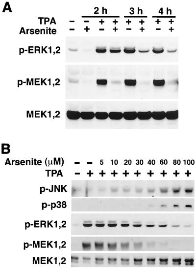
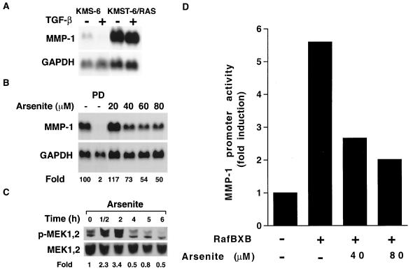
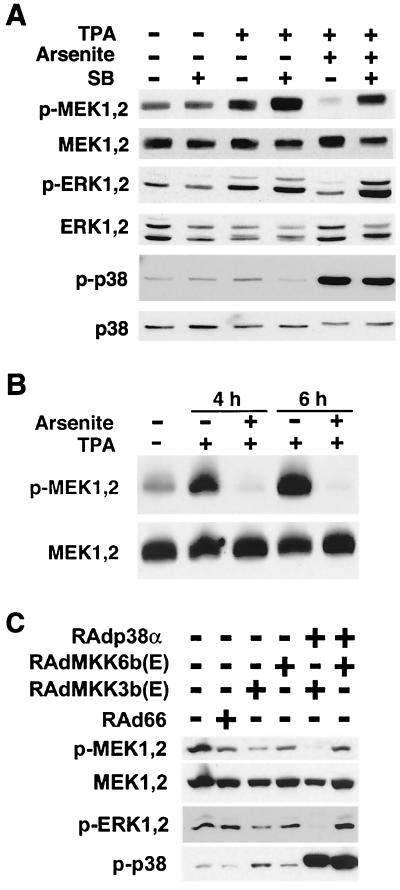
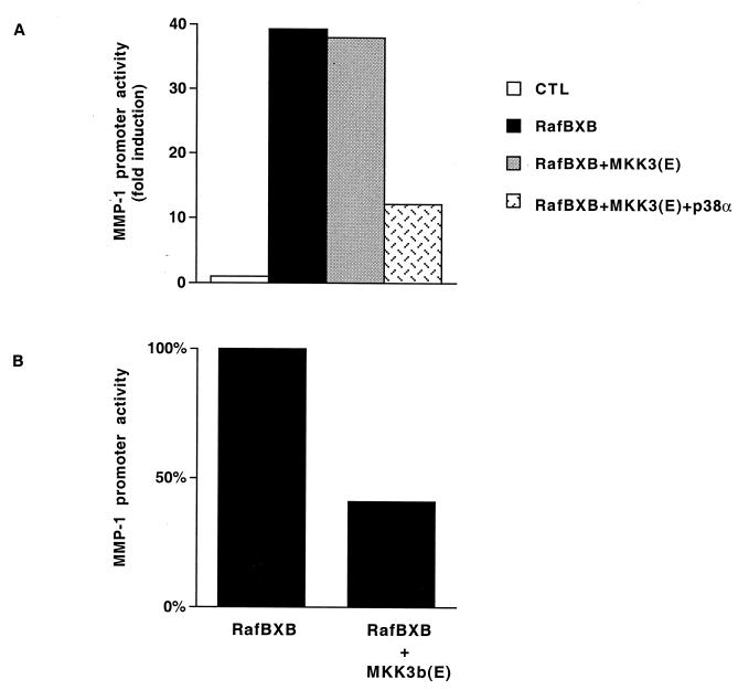
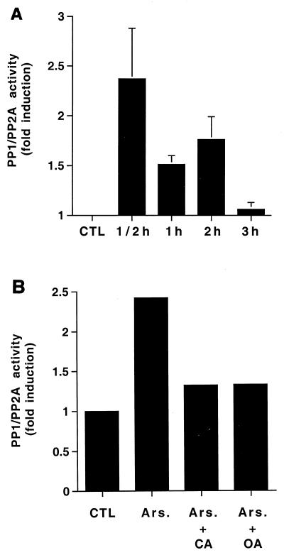
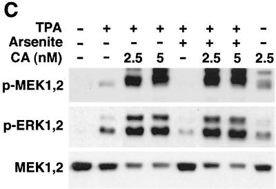
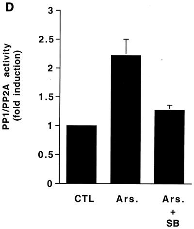
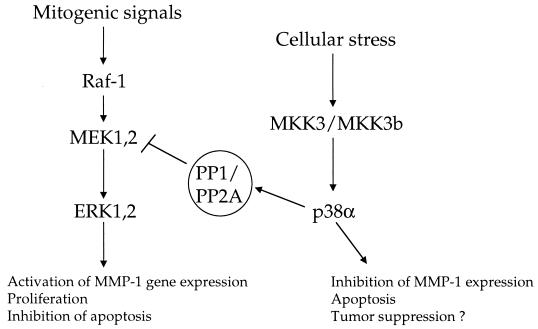
Degradation of collagenous extracellular matrix by collagenase 1 (also known as matrix metalloproteinase 1 [MMP-1]) plays a role in the pathogenesis of various destructive disorders, such as rheumatoid arthritis, chronic ulcers, and tumor invasion and metastasis. Here, we have investigated the role of distinct mitogen-activated protein kinase (MAPK) pathways in the regulation of MMP-1 gene expression. The activation of the extracellular signal-regulated kinase 1 (ERK1)/ERK2 (designated ERK1,2) pathway by oncogenic Ras, constitutively active Raf-1, or phorbol ester resulted in potent stimulation of MMP-1 promoter activity and mRNA expression. In contrast, activation of stress-activated c-Jun N-terminal kinase and p38 pathways by expression of constitutively active mutants of Rac, transforming growth factor beta-activated kinase 1 (TAK1), MAPK kinase 3 (MKK3), or MKK6 or by treatment with arsenite or anisomycin did not alone markedly enhance MMP-1 promoter activity. Constitutively active MKK6 augmented Raf-1-mediated activation of the MMP-1 promoter, whereas active mutants of TAK1 and MKK3b potently inhibited the stimulatory effect of Raf-1. Activation of p38 MAPK by arsenite also potently abrogated stimulation of MMP-1 gene expression by constitutively active Ras and Raf-1 and by phorbol ester. Specific activation of p38alpha by adenovirus-delivered constitutively active MKK3b resulted in potent inhibition of the activity of ERK1,2 and its upstream activator MEK1,2. Furthermore, arsenite prevented phorbol ester-induced phosphorylation of ERK1,2 kinase-MEK1,2, and this effect was dependent on p38-mediated activation of protein phosphatase 1 (PP1) and PP2A. These results provide evidence that activation of signaling cascade MKK3-MKK3b-->p38alpha blocks the ERK1,2 pathway at the level of MEK1,2 via PP1-PP2A and inhibits the activation of MMP-1 gene expression.


Figures

FIG. 1
FIG. 1
Modulation of Raf-1-induced MMP-1 promoter activity by stress-activated MAPK pathways. (A to C) After transfection and glycerol shock, the cultures were maintained for 36 h in 1% FCS, and CAT activity was measured from cell lysates. Transfection efficiency was monitored by cotransfecting the cells with 4 μg of RSV–β-galactoside construct. The values represent the mean of at least two independent experiments, each performed in duplicate. (A) NIH 3T3 fibroblasts were transiently transfected with indicated amounts of the expression vector for the constitutively active form of Raf-1 (RafBXB) together with the -2278CLCAT construct (2 μg), containing 2,278 bp of the human MMP-1 gene promoter linked to the CAT gene. (B) NIH 3T3 fibroblasts were transiently transfected with increasing amounts (0.5, 1, 2, and 4 μg) of expression vectors for constitutively active MKK3 [MKK3(E)], MKK6 [MKK6(EE)], TAK1 (ΔNTAK1), and Rac (RacQL), or with Raf-1 (RafBXB) (2 μg) in combination with MMP-1 promoter-CAT construct -2278CLCAT (2 μg). (C) NIH 3T3 fibroblasts were transiently transfected with MMP-1 promoter-CAT construct -2278CLCAT (1 μg) with the expression vector for constitutively active Raf-1 (RafBXB) alone or in combination with expression vectors for constitutively active MKK3, MKK6, and TAK1 (1 μg each). (D) NIH 3T3 fibroblasts were transiently transfected with 4 μg of expression vectors for the constitutively active form of TAK1 (ΔNTAK1) and Rac [Rac(QL)] together with the expression vector for Flag-tagged p38α. Activity of p38α immunoprecipitated from cell lysates with anti-Flag antibody was determined with GST–ATF-2 as substrate. The levels of p38α in cell lysates were determined by Western blot analysis with anti-Flag antibody. (E) NIH 3T3 fibroblasts were transiently transfected with expression vectors for the constitutively active form of TAK1 (ΔNTAK1) (2 μg), MKK3b [MKK3b(E)] and MKK6b [MKK6b(E)] (4 μg), together with the expression vector for constitutively active Raf-1 (RafBxB) (2 μg). The level of RafBxB in cell lysates was determined by Western blot analysis with c-Raf-1 antibody. (F) NIH 3T3 fibroblasts were transiently transfected with 4 μg of expression vectors for constitutively active forms of MKK3b [MKK3b(E)] and MKK6b [MKK6b(E)], together with expression vectors for wild-type p38 isoforms (p38α, -β, -γ, and -δ) (4 μg each). Control cultures were transfected with the corresponding empty expression vector. Cultures were then maintained for 36 h in 1% FCS–DMEM, and the activity of p38 isoforms immunoprecipitated from cell lysates with anti-Flag antibody was determined using GST–ATF-2 as substrate. CTL, empty expression vector.
FIG. 2
FIG. 2
Arsenite inhibits TPA-elicited induction of MMP-1 expression. Total RNA was isolated and analysed for MMP-1 and GAPDH mRNA abundance by Northern blot hybridizations. Quantitations of MMP-1 mRNA levels corrected for GAPDH mRNA levels are shown below panel C relative to untreated control cultures. (A) Human skin fibroblasts were treated with TPA (60 ng/ml), arsenite (80 μM), and anisomycin (25 ng/ml) alone and in combination for 12 h. (B) Human skin fibroblasts were treated for 12 h with TPA (60 ng/ml) alone or in combination with increasing concentrations of arsenite. (C) HeLa cells were treated with TPA (60 ng/ml) and arsenite (50 μM) alone or in combination for 12 h.
FIG. 3
FIG. 3
Arsenite inhibits activation of ERK1,2. Human skin fibroblasts were treated with arsenite (80 μM) for indicated periods of time. Thereafter, cells were lysed and the levels of phosphorylated ERK1,2 (p-ERK1,2), JNK (p-JNK), and p38 (p-p38) were determined by Western blot analysis with phospho-specific antibodies. Protein loading was studied with antibodies against total p38 and ERK1,2. The cellular levels of catalytic subunits for PP1 and PP2A were determined by Western blot analysis with specific antibodies.
FIG. 4
FIG. 4
Treatment of fibroblasts with arsenite prevents activation of MEK1,2 and ERK1,2. Cells were lysed, and the levels of phosphorylated ERK1,2 (p-ERK1,2), MEK1,2 (p-MEK1,2), JNK (p-JNK), and p-38 (p-p38) were determined by Western blot analysis with corresponding phospho-specific antibodies. Protein loading was determined using antibodies against total MEK1,2. (A) Human skin fibroblasts were treated with TPA (60 ng/ml) alone or in combination with arsenite (80 μM) for indicated periods of time. (B) Human skin fibroblasts were treated with TPA (60 ng/ml) alone or in combination with increasing concentrations of arsenite for 2 h.
FIG. 5
FIG. 5
Arsenite treatment blocks the ERK1,2 pathway downstream of Ras and Raf-1. (A) The Ras-transformed human embryonal fibroblast line (KMST-6/RAS) and the parental fibroblasts (KMS-6) were treated for 24 h with TGF-β (10 ng/ml). The expression of MMP-1 and GAPDH mRNAs was determined by Northern blot hybridizations. (B) KMST-6/RAS cells were incubated for 12 h with MEK1,2 inhibitor PD98059 (PD; 20 μM) or with arsenite in concentrations indicated. Thereafter, expression of MMP-1 and GAPDH mRNAs was determined by Northern blot hybridizations. Quantitation of MMP-1 mRNA levels corrected for GAPDH mRNA levels is shown below the panel relative to untreated control cultures (100). (C) KMST-6/RAS cells were incubated with arsenite (80 μM) for indicated periods of time, and the levels of phosphorylated MEK1,2 (p-MEK1,2) and total MEK1,2 were determined by Western blot analysis. Quantitation of p-MEK1,2 levels is shown below the panel relative to the levels in cultures at time point 0 h (lane 1). (D) Neonatal human skin fibroblasts were transiently transfected with expression vector for the constitutively active form of Raf-1 (RafBXB) together with MMP-1 reporter gene construct -2278CLCAT (2 μg). Twelve hours after the transfection, cells were treated with arsenite (40 or 80 μM), incubation was continued for 12 h, cells were harvested, and CAT activity was measured. The values represent the mean of three experiments each performed in duplicate.
FIG. 6
FIG. 6
Inhibitory effect of arsenite on MEK1,2 activation is mediated by p38 MAPK. (A) Human skin fibroblasts were incubated for 4 h with TPA (60 ng/ml) and arsenite (80 μM) alone or in combination. Where indicated, cells were pretreated for 2 h with specific inhibitor of p38 activity SB203580 (SB; 20 μM). Thereafter, cells were lysed and the levels of phosphorylated MEK1,2 (p-MEK1,2), ERK1,2 (p-ERK1,2), and p38 (p-p38) and total MEK1,2, ERK1,2, and p38 were determined by Western blot analysis. (B) Embryonal fibroblasts from JNK2−/− mice were incubated with TPA (60 ng/ml) alone or in combination with arsenite (80 μM) for the time period indicated. Cells were lysed, and levels of phosphorylated MEK1,2 (p-MEK1,2) and total MEK1,2 were determined by Western blotting. (C) Ras-transformed human embryonal fibroblasts (KMST-6/Ras) were infected with recombinant adenoviruses for constitutively active MKK3b [RAdMKK3b(E)] and MKK6b [RAdMKK6b(E)] alone or in combination with adenovirus harboring wild-type p38α (RAdp38α) at a multiplicity of infection of 500. Control cultures were infected with empty adenovirus RAd66. After 18 h in DMEM with 1% FCS, the medium was changed and the incubations were continued for 48 h. Cells were lysed, and the levels of phosphorylated MEK1,2 (p-MEK1,2), ERK1,2 (p-ERK1,2), and p38 (p-p38) and total MEK1,2 were determined by Western blot analysis.
FIG. 7
FIG. 7
(A) NIH 3T3 fibroblasts were transiently transfected with MMP-1 promoter-reporter gene construct -2278CLCAT (1 μg) and the expression vector for constitutively active Raf-1 (RafBXB) alone or in combination with expression vectors for constitutively active MKK3 [MKK3(E)] and wild-type p38α (1 μg each). Control cultures were transfected with the corresponding empty expression vectors. (B) NIH 3T3 fibroblasts were transiently transfected with -2278CLCAT reporter construct and 1 μg of Raf-1 (RafBXB) alone or in combination with the constitutively active form of MKK3b [MKK3b(E)]. After 36 h, cells were lysed, and CAT activity was measured as an indicator of promoter activity. Transfection efficiency was monitored by cotransfecting the cells with 4 μg of the RSV–β-galactosidase construct. The values represent the mean of two independent experiments each performed in duplicate.
FIG. 8
FIG. 8
Activation of PP1-PP2A is required for arsenite-elicited MEK1,2 dephosphorylation. (A) Human skin fibroblasts were treated with arsenite (80 μM) for the time periods indicated. Thereafter, cells were lysed and equal amounts of protein were assayed for PP1-PP2A phosphatase activity using 32P-labeled glycogen phosphorylase as a substrate. The mean and standard deviation of values from three experiments are shown. x axis, length of arsenite treatment. (B) Human skin fibroblasts were treated with arsenite (Ars; 80 μM) for 30 min. Where indicated, cells were pretreated for 2 h with okadaic acid (OA) (20 ng/ml) or for 15 min with calyculin A (CA) (5 nM). Thereafter, cells were lysed and equal amounts of total protein were assayed for PP1-PP2A activity as described in the legend to panel A. The results of a representative experiment of two experiments with similar results are shown. (C) Human skin fibroblasts were treated for 2 h with TPA (60 ng/ml) and arsenite (80 μM) alone or in combination. Where indicated, cells were pretreated for 15 min with calyculin A (CA) in concentrations shown. The levels of phosphorylated MEK1,2 (p-MEK1,2) and ERK1,2 (p-ERK1,2) were determined by Western blotting using phospho-specific antibodies. The filter was stripped and protein loading was determined by using an antibody against total MEK1,2. A representative blot of two independent experiments with similar results is shown. (D) Human skin fibroblasts were incubated for 30 min with arsenite (Ars; 80 μM). Where indicated cells were pretreated for 2 h with p38 inhibitor SB203580 (SB; 20 μM). Cells were then lysed and PP1-PP2A phosphatase activity was measured as described in the legend to panel A. The mean plus standard deviation of two independent experiments are shown. CTL, untreated control cultures.
FIG. 8
FIG. 8
Activation of PP1-PP2A is required for arsenite-elicited MEK1,2 dephosphorylation. (A) Human skin fibroblasts were treated with arsenite (80 μM) for the time periods indicated. Thereafter, cells were lysed and equal amounts of protein were assayed for PP1-PP2A phosphatase activity using 32P-labeled glycogen phosphorylase as a substrate. The mean and standard deviation of values from three experiments are shown. x axis, length of arsenite treatment. (B) Human skin fibroblasts were treated with arsenite (Ars; 80 μM) for 30 min. Where indicated, cells were pretreated for 2 h with okadaic acid (OA) (20 ng/ml) or for 15 min with calyculin A (CA) (5 nM). Thereafter, cells were lysed and equal amounts of total protein were assayed for PP1-PP2A activity as described in the legend to panel A. The results of a representative experiment of two experiments with similar results are shown. (C) Human skin fibroblasts were treated for 2 h with TPA (60 ng/ml) and arsenite (80 μM) alone or in combination. Where indicated, cells were pretreated for 15 min with calyculin A (CA) in concentrations shown. The levels of phosphorylated MEK1,2 (p-MEK1,2) and ERK1,2 (p-ERK1,2) were determined by Western blotting using phospho-specific antibodies. The filter was stripped and protein loading was determined by using an antibody against total MEK1,2. A representative blot of two independent experiments with similar results is shown. (D) Human skin fibroblasts were incubated for 30 min with arsenite (Ars; 80 μM). Where indicated cells were pretreated for 2 h with p38 inhibitor SB203580 (SB; 20 μM). Cells were then lysed and PP1-PP2A phosphatase activity was measured as described in the legend to panel A. The mean plus standard deviation of two independent experiments are shown. CTL, untreated control cultures.
FIG. 9
FIG. 9
Schematic presentation of the proposed opposite roles of ERK1,2 and MKK3/MKK3b→p38α pathways in the regulation of cell behavior.

Similar articles

Cited by

References

    1. Abraham D, Podar K, Pacher M, Kubicek M, Welzel N, Hemmings B A, Dilworth S M, Mischak H, Kolch W, Baccarini M. Raf-1-associated PP2A as a positive regulator of kinase activation. J Biol Chem. 2000;275:22300–22304. - PubMed
    1. Bruder J T, Heidecker G, Rapp U R. Serum, TPA-, and Ras-induced expression from Ap-1/Ets driven promoters requires Raf-1 kinase. Genes Dev. 1992;6:545–556. - PubMed
    1. Casillas A M, Amaral K, Chegini Farahani S, Nel A E. Okadaic acid activates p42 mitogen-activated protein kinase (MAP kinase; ERK-2) in B-lymphocytes but inhibits rather than augments cellular proliferation: contrast with phorbol 12-myristate 13-acetate. Biochem J. 1993;290:545–550. - PMC - PubMed
    1. Cavigelli M, Li W W, Lin A, Su B, Yoshioka K, Karin M. The tumor promoter arsenite stimulates AP-1 activity by inhibiting a JNK phosphatase. EMBO J. 1996;15:6269–6279. - PMC - PubMed
    1. Chen Y, Lin-Shiau S, Lin J. Involvement of reactive oxygen species and caspase 3 activation in arsenite-induced apoptosis. J Cell Physiol. 1998;177:324–333. - PubMed

Publication types

MeSH terms