. 2001 Apr;21(7):2404-12.
doi: 10.1128/MCB.21.7.2404-2412.2001.

Molecular distinction between specification and differentiation in the myogenic basic helix-loop-helix transcription factor family

Affiliations

Molecular distinction between specification and differentiation in the myogenic basic helix-loop-helix transcription factor family

D A Bergstrom et al. Mol Cell Biol. 2001 Apr.

Abstract

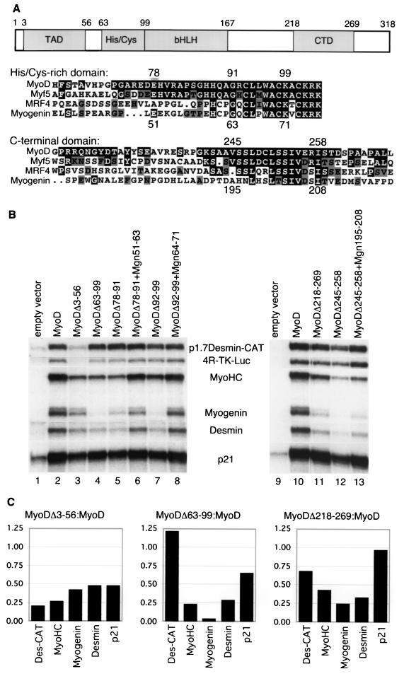
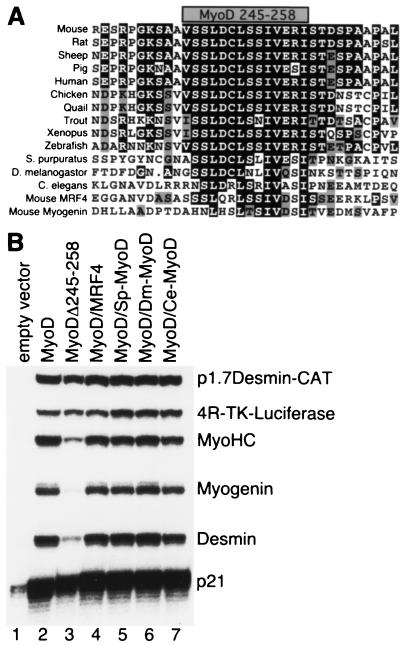
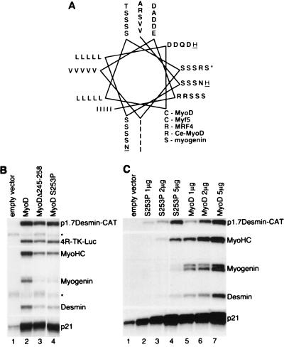
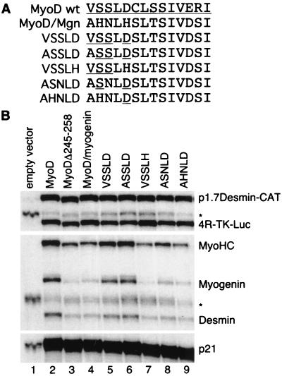
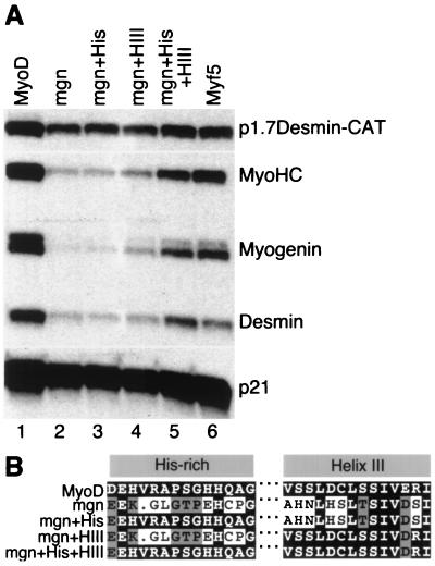
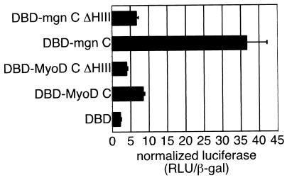
The myogenic basic helix-loop-helix (bHLH) proteins regulate both skeletal muscle specification and differentiation: MyoD and Myf5 establish the muscle lineage, whereas myogenin mediates differentiation. Previously, we demonstrated that MyoD was more efficient than myogenin at initiating the expression of skeletal muscle genes, and in this study we present the molecular basis for this difference. A conserved amphipathic alpha-helix in the carboxy terminus of the myogenic bHLH proteins has distinct activities in MyoD and myogenin: the MyoD helix facilitates the initiation of endogenous gene expression, whereas the myogenin helix functions as a general transcriptional activation domain. Thus, the alternate use of a similar motif for gene initiation and activation provides a molecular basis for the distinction between specification and differentiation within the myogenic bHLH gene family.


Figures

FIG. 1
FIG. 1
The functional differences between MyoD and myogenin map to a C-terminal domain. (A) Schematic diagram of MyoD domains and partial sequence alignment of the murine myogenic bHLH proteins, corresponding to MyoD amino acids 63 to 104 and 218 to 269. MyoD amino acid numbers are indicated along the top, and myogenin amino acid numbers are along the bottom of each alignment. (B) S1 nuclease protection of RNA from NIH 3T3 cells transiently transfected with expression vectors for MyoD or MyoD mutants, as indicated. Protected messages are identified between the two gel images. When normalized to p1.7Des-Cat, MyoDΔ63-99 (lane 4) was 3% as efficient as MyoD at initiating expression of the endogenous myogenin gene, 24% as efficient on the endogenous desmin gene, and 20% as efficient on the endogenous myosin heavy chain gene; it was nearly equal to MyoD at increasing expression from the preinitiated p21 gene (81% relative to MyoD). Each mutant was analyzed multiple times, and the figure represents a typical experiment. (C) Ratio of the signal in select mutants relative to wild-type MyoD. Deletion of the activation domain (MyoDΔ3-56) results in a relatively equal decrease in the expression level of all of the target genes. In comparison, the MyoDΔ63-99 and MyoDΔ218-269 deletions show relatively preserved activity on the p1.7Des-CAT reporter and the endogenous p21 gene compared to their activity on the endogenous MyoHC, myogenin, and desmin genes.
FIG. 2
FIG. 2
Conservation of the function of the MyoD C-terminal motif. (A) Multiple sequence alignment of MyoD proteins from several vertebrate and invertebrate species and from murine MRF4 and myogenin in the region corresponding to the murine MyoD C-terminal domain. The shaded box at the top of the alignment represents the critical motif encoded by amino acids 245 to 258. (B) S1 protection of RNA following transfection of NIH 3T3 cells with MyoD wild-type (MyoD) or chimeric proteins as indicated, showing functional conservation of the MyoD C-terminal domain. For substitution mutants, the sequence aligning with murine MyoD 245 to 258 in panel A was substituted for the murine MyoD sequence to generate chimeric proteins, as follows: MyoD/MRF4, murine MRF4 sequence; MyoD/Sp-MyoD, S. purpuratus MyoD sequence; MyoD/Dm-MyoD, D. melanogaster MyoD sequence; and MyoD/Ce-MyoD, C. elegans MyoD sequence. Chimeric proteins were generated by ligating oligonucleotides encoding the desired C-terminal domain into a murine MyoD shuttle vector, from which the wild-type C-terminal domain was removed and replaced with an NheI site.
FIG. 3
FIG. 3
The function of the carboxy-terminal domain of the myogenic bHLH proteins requires the ability to adopt an amphipathic alpha-helical conformation. (A) Helical wheel representation of the C-terminal motif aligning murine MyoD, Myf5, MRF4, C. elegans MyoD, and murine myogenin, demonstrating the high degree of conservation of the hydrophobic face of the helix and the more variable hydrophilic face. (B) S1 nuclease protection of RNA from NIH 3T3 fibroblasts transfected with wild-type MyoD (MyoD), MyoDΔ245-58, and MyoD-S253P as indicated. Serine-253 (indicated by ∗ in panel A) falls in the middle of the predicted helix on the hydrophilic face. (C) Titration of the MyoD-S253P mutant compared to wild-type MyoD. The indicated amounts of expression plasmid were transfected. Empty expression vector was used to adjust the total quantity of transfected plasmid.
FIG. 4
FIG. 4
Residues on the hydrophilic face of helix III differentiate MyoD and myogenin function. (A) The MyoDΔ245-258–myogenin 195-208 chimera was subjected to back mutagenesis, represented in this schematic. MyoD residues are underlined. Back mutants are identified by the sequence of the first five amino acids of helix III, i.e., VSSLD represents substitution of MyoD amino acids 245 to 249 into the chimeric protein. (B) S1 nuclease protection analysis of back mutations in the chimeric MyoDΔ245-258–myogenin 195-208 protein. ∗, partially digested probe.
FIG. 5
FIG. 5
Role of putative phosphorylation sites in helix III regulation. Serine residues at MyoD positions 246 and 253 were replaced with alanine or glutamate residues. The ability of the mutants to initiate expression of endogenous skeletal muscle genes was assayed using an S1 nuclease protection assay. ∗, partially digested probe.
FIG. 6
FIG. 6
MyoD helix III (HIII) increases the ability of myogenin to efficiently initiate expression of skeletal muscle genes. (A) MyoD helix III increases the ability of myogenin to efficiently initiate expression of skeletal muscle genes. S1 nuclease assay of RNA from NIH 3T3 cells transfected with wild-type MyoD (lane 1), wild-type myogenin (lane 2), myogenin-MyoD chimeras (lanes 3 to 6), and Myf5 (lane 7). (B) Alignment of MyoD, myogenin, and the chimeric proteins: mgn+His was constructed by substituting 10 amino acids from MyoD into the corresponding myogenin region; mgn+HIII required 7 amino acid substitutions.
FIG. 7
FIG. 7
Myogenin possess a helix III-dependent C-terminal activation domain. The C termini of MyoD and myogenin were assayed for the ability to function as a general activation domain when fused to the Gal4 DBD (amino acids 1 to 147). Gal fusion proteins were generated by PCR amplifying cDNA encoding MyoD amino acids 170 to 318 (DBD-MyoD C) and myogenin amino acids 136 to 224 (DBD-mgn C) and ligating the PCR products into the pSG424 vector. Similar fusion proteins with deletions of the helix III motif in MyoD (amino acids 245 to 258; DBD-MyoD C ΔHIII) and myogenin (amino acids 195 to 208; DBD-mgn C ΔHIII) were also made. The Gal4-C terminus chimeras were cotransfected into NIH 3T3 cells with a Gal4-responsive luciferase reporter plasmid and a constitutively expressed β-galactosidase reporter to normalize for transfection efficiency. Each transfection was repeated three times. The graph represents the mean normalized luciferase activity (relative light units [RLU]/β-galactosidase activity) for the three experiments. Error bars represent standard deviations.

References

    1. Braun T, Buschhausen-Denker G, Bober E, Tannich E, Arnold H H. A novel human muscle factor related to but distinct from MyoD1 induces myogenic conversion in 10T1/2 fibroblasts. EMBO J. 1989;8:701–709. - PMC - PubMed
    1. Edmondson D G, Olson E N. A gene with homology to the myc similarity region of MyoD1 is expressed during myogenesis and is sufficient to activate the muscle differentiation program. Genes Dev. 1989;3:628–640. . (Erratum, 4:1450, 1990.) - PubMed
    1. Fujisawa-Sehara A, Nabeshima Y, Hosoda Y, Obinata T. Myogenin contains two domains conserved among myogenic factors. J Biol Chem. 1990;265:15219–15223. - PubMed
    1. Gaudreau L, Schmid A, Blaschke D, Ptashne M, Horz W. RNA polymerase II holoenzyme recruitment is sufficient to remodel chromatin at the yeast PHO5 promoter. Cell. 1997;89:55–62. - PubMed
    1. Gerber A N, Klesert T R, Bergstrom D A, Tapscott S J. Two domains of MyoD mediate transcriptional activation of genes in repressive chromatin: a mechanism for lineage determination in myogenesis. Genes Dev. 1997;11:436–450. - PubMed

Publication types

LinkOut - more resources