. 2001 Apr;21(7):2449-62.
doi: 10.1128/MCB.21.7.2449-2462.2001.

The Cbk1p pathway is important for polarized cell growth and cell separation in Saccharomyces cerevisiae

Affiliations

The Cbk1p pathway is important for polarized cell growth and cell separation in Saccharomyces cerevisiae

S Bidlingmaier et al. Mol Cell Biol. 2001 Apr.

Abstract

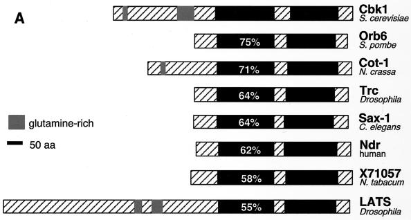
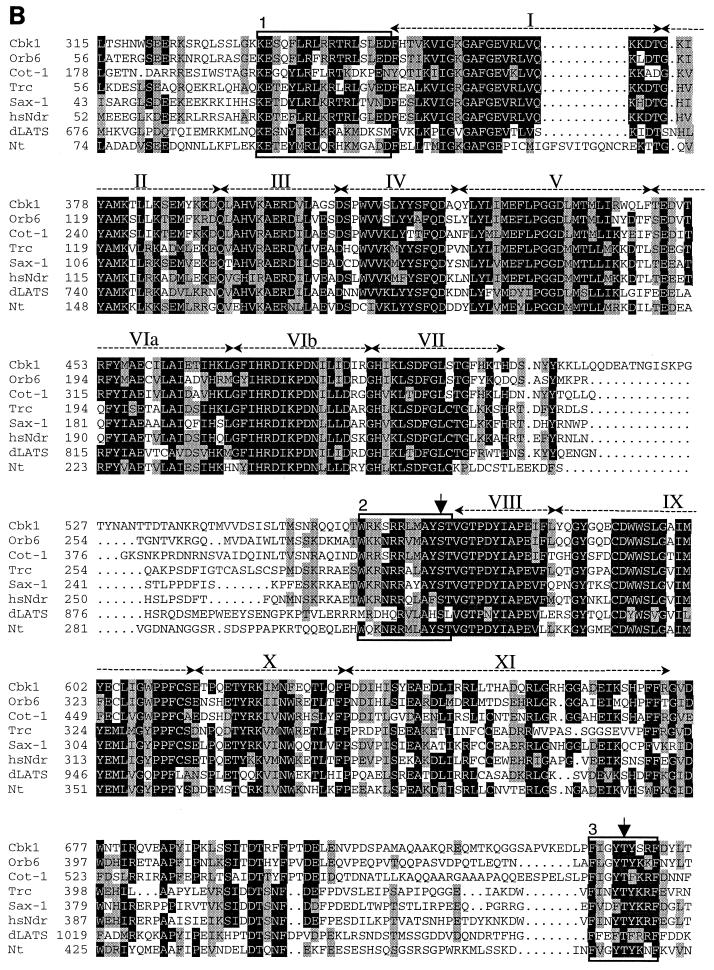
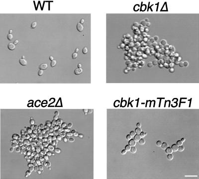
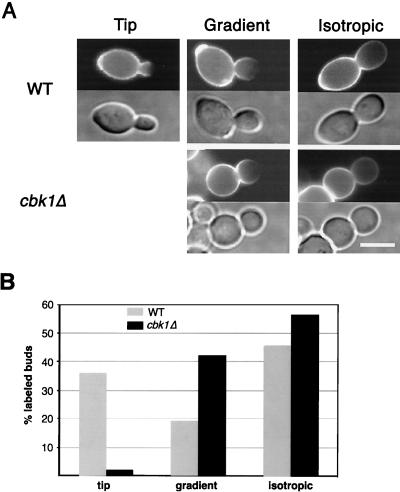
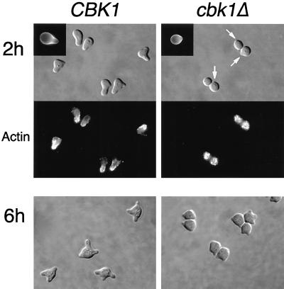
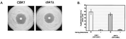
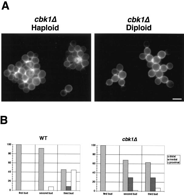
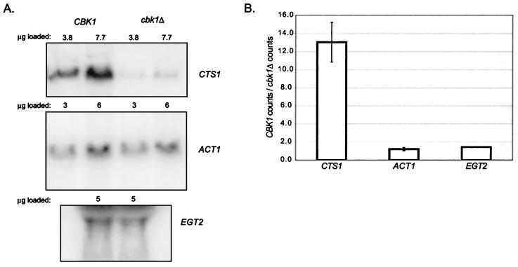
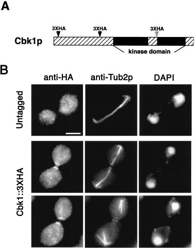
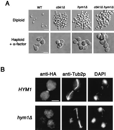
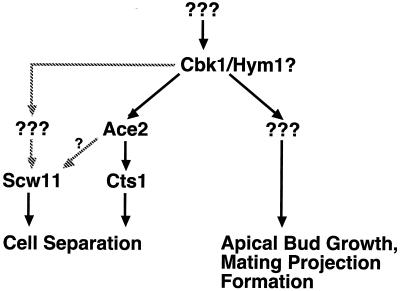
During the early stages of budding, cell wall remodeling and polarized secretion are concentrated at the bud tip (apical growth). The CBK1 gene, encoding a putative serine/threonine protein kinase, was identified in a screen designed to isolate mutations that affect apical growth. Analysis of cbk1Delta cells reveals that Cbk1p is required for efficient apical growth, proper mating projection morphology, bipolar bud site selection in diploid cells, and cell separation. Epitope-tagged Cbk1p localizes to both sides of the bud neck in late anaphase, just prior to cell separation. CBK1 and another gene, HYM1, were previously identified in a screen for genes involved in transcriptional repression and proposed to function in the same pathway. Deletion of HYM1 causes phenotypes similar to those observed in cbk1Delta cells and disrupts the bud neck localization of Cbk1p. Whole-genome transcriptional analysis of cbk1Delta suggests that the kinase regulates the expression of a number of genes with cell wall-related functions, including two genes required for efficient cell separation: the chitinase-encoding gene CTS1 and the glucanase-encoding gene SCW11. The Ace2p transcription factor is required for expression of CTS1 and has been shown to physically interact with Cbk1p. Analysis of ace2Delta cells reveals that Ace2p is required for cell separation but not for polarized growth. Our results suggest that Cbk1p and Hym1p function to regulate two distinct cell morphogenesis pathways: an ACE2-independent pathway that is required for efficient apical growth and mating projection formation and an ACE2-dependent pathway that is required for efficient cell separation following cytokinesis. Cbk1p is most closely related to the Neurospora crassa Cot-1; Schizosaccharomyces pombe Orb6; Caenorhabditis elegans, Drosophila, and human Ndr; and Drosophila and mammalian WARTS/LATS kinases. Many Cbk1-related kinases have been shown to regulate cellular morphology.


Figures

FIG. 1
FIG. 1
Morphology of cdc34-2 and cdc34-2 cbk1Δ cells grown at the restrictive temperature. Cells were grown to mid-logarithmic phase at 25°C in YPAD, shifted to 37°C for 7 h, and then fixed with formaldehyde and photographed. Bar, 5.0 μm.
FIG. 2
FIG. 2
Sequence comparison of Cbk1p-related protein kinases. (A) The predicted amino acid (aa) sequences of Cbk1p from S. cerevisiae, Orb6 from S. pombe (53), Cot-1 from N. crassa (55), human Ndr (34), Drosophila Trc (Ndr) (15), C. elegans SAX-1 (Ndr) (56), N. tabacum kinase X71057 (GenBank accession no. X71057), and Drosophila melanogaster LATS (21, 54) were compared using the program CLUSTALW (48). Kinase domains (19) are shown in black, and the percent amino acid identity of the kinase domains with the Cbk1p kinase domain is shown in white. Regions outside the putative catalytic domain are crosshatched. The insert between subdomains VII and VIII of the catalytic domain (crosshatched region in the middle of the kinase domain) is characteristic of this family of protein kinases. Several members of this kinase family also have glutamine-rich regions in the sequence N terminal of their catalytic domains. (B) Multiple-sequence alignment of the catalytic domain and surrounding sequences of the Cbk1p-related kinases was performed using CLUSTALW. Shading was done using MacBoxShade (M. Baron). Shaded residues are conserved in at least five of the eight sequences. Black shading indicates identity with Cbk1p, while grey shading indicates similarity. Kinase subdomains are labeled above the sequence in roman numerals. Numbered boxed sequences indicate functionally significant regions discussed in the text. Boxed region 1, directly preceding the kinase domain, overlaps the Ca2+/S100B-binding domain of Ndr. Boxed region 2, directly preceding kinase subdomain VIII, indicates a conserved motif that overlaps the activation loop phosphorylation site in human Ndr. Boxed region 3, near the carboxy terminus, corresponds to the hydrophobic motif that is found in many AGC family kinases. Residues corresponding to experimentally determined sites of regulatory phosphorylation in human Ndr are marked with arrows.
FIG. 2
FIG. 2
Sequence comparison of Cbk1p-related protein kinases. (A) The predicted amino acid (aa) sequences of Cbk1p from S. cerevisiae, Orb6 from S. pombe (53), Cot-1 from N. crassa (55), human Ndr (34), Drosophila Trc (Ndr) (15), C. elegans SAX-1 (Ndr) (56), N. tabacum kinase X71057 (GenBank accession no. X71057), and Drosophila melanogaster LATS (21, 54) were compared using the program CLUSTALW (48). Kinase domains (19) are shown in black, and the percent amino acid identity of the kinase domains with the Cbk1p kinase domain is shown in white. Regions outside the putative catalytic domain are crosshatched. The insert between subdomains VII and VIII of the catalytic domain (crosshatched region in the middle of the kinase domain) is characteristic of this family of protein kinases. Several members of this kinase family also have glutamine-rich regions in the sequence N terminal of their catalytic domains. (B) Multiple-sequence alignment of the catalytic domain and surrounding sequences of the Cbk1p-related kinases was performed using CLUSTALW. Shading was done using MacBoxShade (M. Baron). Shaded residues are conserved in at least five of the eight sequences. Black shading indicates identity with Cbk1p, while grey shading indicates similarity. Kinase subdomains are labeled above the sequence in roman numerals. Numbered boxed sequences indicate functionally significant regions discussed in the text. Boxed region 1, directly preceding the kinase domain, overlaps the Ca2+/S100B-binding domain of Ndr. Boxed region 2, directly preceding kinase subdomain VIII, indicates a conserved motif that overlaps the activation loop phosphorylation site in human Ndr. Boxed region 3, near the carboxy terminus, corresponds to the hydrophobic motif that is found in many AGC family kinases. Residues corresponding to experimentally determined sites of regulatory phosphorylation in human Ndr are marked with arrows.
FIG. 3
FIG. 3
Morphology of wild-type (WT) and cbk1Δ, ace2Δ, and cbk1-mTn3F1 mutant homozygous diploid cells. Cells were grown to early logarithmic phase in YPAD and then fixed with formaldehyde and photographed. Bar, 10 μm.
FIG. 4
FIG. 4
Electron microscopy of high-pressure-frozen–freeze-substituted cbk1Δ cells. Black arrowheads indicate regions of shared cell wall (CW) material. No cytoplasmic connections were observed in adjacent sections at any of these sites. Bar, 0.5 μm.
FIG. 5
FIG. 5
Analysis of apical growth in wild-type (WT) and cbk1Δ mutant cells by FITC-ConA pulse-labeling. (A) Exponentially growing wild-type and cbk1Δ mutant diploid cells were pulse-labeled with FITC-ConA for 10 min, washed, and returned to growth in fresh medium. After 25 min, cells were fixed and observed by fluorescence microscopy. FITC-ConA fluorescence images are shown above differential interference contrast images. Staining that is completely faded at the bud tip indicates apical growth (Tip), while uniformly decreased staining throughout the bud indicates isotropic growth (Isotropic). Cells with a gradient of staining that was not completely faded at the bud tip were also observed (Gradient). (B) The pattern of fading in labeled buds was quantitated for wild-type and cbk1Δ diploid cells. Labeled buds were divided into three categories as described above: labeled buds with a completely faded tip (tip), labeled buds with staining that faded toward the tip but with staining at the tip still visible (gradient), and labeled buds with uniformly faded staining (isotropic). Bar, 5.0 μm.
FIG. 6
FIG. 6
Pheromone-induced morphology and actin reorganization in wild-type and cbk1Δ mutant cells. Exponentially growing cells were incubated with α-factor (5 μg/ml) for 2 or 6 h, fixed with formaldehyde, and stained with rhodamine-phalloidin to visualize the actin cytoskeleton. White arrows point out small protrusions on the cell surface. Upper left insets show calcofluor staining of chitin. Strains used for the 6-h time point were bar1Δ mutants. Bar, 5.0 μm.
FIG. 7
FIG. 7
Pheromone-induced growth arrest and transcriptional induction in wild-type and cbk1Δ mutant cells. (A) Sterile filter disks were saturated with α-factor at 5 μg/ml and placed in the middle of freshly plated lawns of wild-type or cbk1Δ mutant cells. Pictures were taken after 2 days of incubation at 30°C. The strains used were bar1Δ mutants. (B) Expression of FUS1-lacZ reporter fusion upon exposure to α-factor at 5 μg/ml in YPAD medium. Open bars represent wild-type cells; gray bars represent cbk1Δ mutant cells. β-Galactosidase expression levels were measured in Miller units as previously described (55).
FIG. 8
FIG. 8
Bud site selection in wild-type (WT) and cbk1Δ mutant cells. (A) Bud scars in haploid and diploid cbk1Δ cells were stained with calcofluor and photographed. Note the chitin-rich junctions between attached cells. Bar, 5.0 μm. (B) The graphs show the placement of the first three buds in wild-type and cbk1Δ mutant diploid cells. Bud scars were scored as distal (opposite the birth site), proximal (near the birth site), or medial (see Materials and Methods). At least 100 cells of each type (first, second, and third buds) were analyzed for both strains.
FIG. 9
FIG. 9
Northern analysis of CTS1 mRNA levels in wild-type and cbk1Δ mutant cells. (A) Northern blots showing CTS1 (top), ACT1 (middle), and EGT2 (bottom) message levels in wild-type and cbk1Δ mutant cells. The amount of total RNA loaded in each lane is indicated above the corresponding lane. (B) Relative expression of CTS1, ACT1, and EGT2 mRNAs in wild-type and cbk1Δ cells, expressed as the ratio of PhosphorImager counts (arbitrary units) of mRNA from wild-type cells over mRNA from cbk1Δ mutant cells.
FIG. 10
FIG. 10
Localization of Cbk1p by indirect immunofluorescence staining. (A) The chromosomal CBK1 gene was tagged with a triple HA epitope (3xHA) at the indicated positions (see Materials and Methods). Dark arrowheads indicate that the tagged protein retained its function, and the light arrowhead represents the cbk1-mTn3F1 allele, which is partially functional (details are discussed in Results). (B) CBK1::3xHA and untagged haploid strains were stained with anti-HA antibodies, anti-Tub2p antibodies (microtubule staining), and DAPI (DNA staining). Cbk1p::3xHA localizes to both sides of the bud neck late in anaphase (note long spindle length) and remains as a patch on the surface of each recently separated cell. No polarized localization was detected in the untagged strain. For these pictures, the strain with CBK1 tagged at amino acid 259 was used. Bar, 5.0 μm.
FIG. 11
FIG. 11
hym1Δ and cbk1Δ mutants have similar phenotypes, and Hym1p is required for normal Cbk1p localization. (A) Morphology of vegetatively growing and pheromone-treated wild-type (WT) and cbk1Δ, hym1Δ, and cbk1Δ hym1Δ mutant cells. Exponentially growing diploid cells were fixed with formaldehyde and photographed. Bar, 10 μm. Exponentially growing haploid cells were treated with α-factor at 5 μg/ml for 2 h, fixed, and then photographed. Bar, 5.0 μm. (B) Cbk1p::3xHA was visualized by indirect immunofluorescence assay in wild-type and hym1Δ mutant cells. No polarized localization of Cbk1p::3xHA is observed in hym1Δ mutant cells. Bar, 5.0 μm.
FIG. 12
FIG. 12
Model for the regulation of morphogenesis by Cbk1p and Hym1p. Cbk1p functions in two distinct cell morphogenesis pathways: an ACE2-independent pathway that is required for apical growth and mating projection formation and an ACE2-dependent pathway that is required for cell separation. Based on genetic analysis, Hym1p is proposed to act at the same level as Cbk1p. Expression of SCW11 may be Ace2p dependent or require another, unknown, transcription factor. Downstream targets of Cbk1p that promote apical growth and mating projection formation await identification.

References

    1. Ausubel F, Brent R, Kingston R, Moore D, Seidman J, Smith J, Struhl K, editors. Current protocols in molecular biology. New York, N.Y: John Wiley & Sons, Inc.; 2000.
    1. Baudin A, Ozier-Kalogeropoulos O, Denouel A, Lacroute F, Cullin C. A simple and efficient method for direct gene deletion in Saccharomyces cerevisiae. Nucleic Acids Res. 1993;21:3329–3330. - PMC - PubMed
    1. Bedinger P A, Hardeman K J, Loukides C A. Travelling in style: the cell biology of pollen. Trends Cell Biol. 1994;4:132–138. - PubMed
    1. Belham C, Wu S, Avruch J. Intracellular signalling: PDK1—a kinase at the hub of things. Curr Biol. 1999;9:R93–R96. - PubMed
    1. Bulawa C E, Slater M, Cabib E, Au-Young J, Sburlati A, Adair W L, Jr, Robbins P W. The S. cerevisiae structural gene for chitin synthase is not required for chitin synthesis in vivo. Cell. 1986;46:213–225. - PubMed

Publication types

MeSH terms

Substances